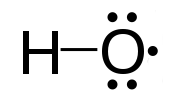
Photo from wikipedia
Total redox capacity (TRC) and oxidative stress (OxiStress) of biological objects (such as cells, tissues, and body fluids) are some of the most frequently analyzed parameters in life science. Development… Click to show full abstract
Total redox capacity (TRC) and oxidative stress (OxiStress) of biological objects (such as cells, tissues, and body fluids) are some of the most frequently analyzed parameters in life science. Development of highly sensitive molecular probes and analytical methods for detection of these parameters is a rapidly growing sector of BioTech's R&D industry. The aim of the present study was to develop quantum sensors for tracking the TRC and/or OxiStress in living biological objects using electron-paramagnetic resonance (EPR), magnetic resonance imaging (MRI), and optical imaging. We describe a two-set sensor system: (i) TRC sensor QD@CD-TEMPO and (ii) OxiStress sensor QD@CD-TEMPOH. Both redox sensors are composed of small-size quantum dots (QDs), coated with multinitroxide-functionalized cyclodextrin (paramagnetic CD-TEMPO or diamagnetic CD-TEMPOH) conjugated with triphenylphosphonium (TPP) groups. The TPP groups were added to achieve intracellular delivery and mitochondrial localization. Nitroxide residues interact simultaneously with various oxidizers and reducers, and the sensors are transformed from the paramagnetic radical form (QD@CD-TEMPO) into diamagnetic hydroxylamine form (QD@CD-TEMPOH) and vice-versa, because of nitroxide redox-cycling. These chemical transformations are accompanied by characteristic dynamics of their contrast features because of quenching of QD fluorescence by nitroxide radicals. The TRC sensor was applied for EPR analysis of cellular redox-status in vitro on isolated cells with different proliferative indexes, as well as for noninvasive MRI of redox imbalance and severe oxidative stress in vivo on mice with renal dysfunction.
Journal Title: Analytical chemistry
Year Published: 2021
Link to full text (if available)
Share on Social Media: Sign Up to like & get
recommendations!