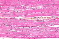
Photo from wikipedia
The potential of electrospun polydioxanone (PDX) mats as scaffolds for skeletal tissue regeneration was significantly enhanced through improvement of the cell-mediated biomimetic mineralization and multicellular response. This was achieved by… Click to show full abstract
The potential of electrospun polydioxanone (PDX) mats as scaffolds for skeletal tissue regeneration was significantly enhanced through improvement of the cell-mediated biomimetic mineralization and multicellular response. This was achieved by blending PDX ( i) with poly(hydroxybutyrate- co-valerate) (PHBV) in the presence of hydroxyapatite (HA) and ( ii) with aloe vera (AV) extract containing a mixture of acemannan/glucomannan. In an exhaustive study, the behavior of the most relevant cell lines involved in the skeletal tissue healing cascade, i.e. fibroblasts, macrophages, endothelial cells and preosteoblasts, on the scaffolds was investigated. The scaffolds were shown to be nontoxic, to exhibit insignificant inflammatory responses in macrophages, and to be degradable by macrophage-secreted enzymes. As a result of different phase separation in PDX/PHBV/HA and PDX/AV blend mats, cells interacted differentially. Presumably due to varying tension states of cell-matrix interactions, thinner microtubules and significantly more cell adhesion sites and filopodia were formed on PDX/AV compared to PDX/PHBV/HA. While PDX/PHBV/HA supported micrometer-sized spherical particles, nanosized rod-like HA was observed to nucleate and grow on PDX/AV fibers, allowing the mineralized PDX/AV scaffold to retain its porosity over a longer time for cellular infiltration. Finally, PDX/AV exhibited better in vivo biocompatibility compared to PDX/PHBV/HA, as indicated by the reduced fibrous capsule thickness and enhanced blood vessel formation. Overall, PDX/AV blend mats showed a significantly enhanced potential for skeletal tissue regeneration compared to the already promising PDX/PHBV/HA blends.
Journal Title: ACS applied materials & interfaces
Year Published: 2019
Link to full text (if available)
Share on Social Media: Sign Up to like & get
recommendations!