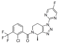
Photo from wikipedia
Radioligands targeting microglia cells have been developed to identify and determine neuroinflammation in the living brain. One recently discovered ligand is JNJ-64413739 that binds selectively to the purinergic receptor P2X7R.… Click to show full abstract
Radioligands targeting microglia cells have been developed to identify and determine neuroinflammation in the living brain. One recently discovered ligand is JNJ-64413739 that binds selectively to the purinergic receptor P2X7R. The expression of P2X7R is increased under inflammation; hence, the ligand is considered useful in the detection of neuroinflammation in the brain. [18F]JNJ-64413739 has been evaluated in healthy subjects with positron emission tomography; however, the in vitro binding properties of the ligand in human brain tissue have not been investigated. Therefore, the purpose of this study was to measure Bmax and Kd of [3H]JNJ-64413739 using autoradiography on human cortical tissue sections resected from a total of 48 patients with treatment-resistant epilepsy. Correlations between the specific binding of [3H]JNJ-64413739 with age, sex, and duration of disease were explored. Finally, to examine the relationship between P2X7R and TSPO availability, specific binding of [3H]JNJ-64413739 and [123I]CLINDE was examined in the same tissue. The binding was measured in both cortical gray and subcortical white matter. Saturation revealed a Kd (5 nM) value similar between gray and white matter but a larger Bmax in the white than in the gray matter. The binding was completely displaced by the cold ligand and structurally different P2X7R ligands. The variability in saturable binding among the samples was found to be 38% in gray and white matter but was not correlated to either age, sex, or the duration of the disease. Interestingly, there was no significant correlation between [3H]JNJ-64413739 and [123I]CLINDE binding. These data demonstrate that [3H]JNJ-64413739 is a suitable radioligand for evaluating the distribution and expression of the P2X7R in the human brain.
Journal Title: ACS Chemical Neuroscience
Year Published: 2022
Link to full text (if available)
Share on Social Media: Sign Up to like & get
recommendations!