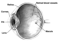
Photo from wikipedia
PurposeTo describe optical coherence tomography (OCT) features in the Bruch’s membrane (BM) of eyes with angioid streaks (AS) and evaluate their evolution over the follow-up.Patients and methodsPatients with AS presenting… Click to show full abstract
PurposeTo describe optical coherence tomography (OCT) features in the Bruch’s membrane (BM) of eyes with angioid streaks (AS) and evaluate their evolution over the follow-up.Patients and methodsPatients with AS presenting between March 2016 and September 2016 at two tertiary referral centers were consecutively recruited in this study. Eligibility criteria included prior spectral domain (SD)-OCT images, taken at least 3 months before at the same referral center, with automated eye tracking and image alignment modules. Alterations of BM were described and compared to previous scans over the follow-up. Multimodal imaging was used to identify alteration of retinal pigment epithelium (RPE) and choroid.ResultsThirty-two eyes of 16 consecutive patients with AS were included. BM undulations, mostly observed around the optic nerve head, were found in 19 (59.4%) of 32 eyes. BM breaks were found in 31 (96.9%) out of 32 eyes. Evolution of BM undulations into BM breaks was observed in 5 eyes (15.6%). Choroidal neovascularization (CNV) was observed in 12 eyes (37.5%) during follow-up, typically in areas of BM interruption.ConclusionsBM undulations, probably caused by high stretching forces exerted on the BM around the optic nerve head, seem to precede some BM breaks. BM interruptions may be a preferred way for the growth of CNV, which was identified in one-third of our cases.
Journal Title: Eye
Year Published: 2017
Link to full text (if available)
Share on Social Media: Sign Up to like & get
recommendations!