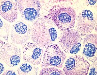
Photo from wikipedia
Targeted functional genomics represents a powerful approach for studying gene function in vivo and in vitro. However, its application to gene expression studies in human mast cells has been hampered… Click to show full abstract
Targeted functional genomics represents a powerful approach for studying gene function in vivo and in vitro. However, its application to gene expression studies in human mast cells has been hampered by low yields of human mast cell cultures and their poor transfection efficiency. We developed an imaging system in which mast cell degranulation can be visualized in single cells subjected to shRNA knockdown or CRISPR–Cas9 gene editing. By using high-resolution confocal microscopy and a fluorochrome-labeled avidin probe, one can directly assess the alteration of functional responses, i.e., degranulation, in single human mast cells (10–12 weeks old). The elimination of a drug or marker selection step avoids the use of potentially toxic treatment procedures, and the brief hands-on time of the functional analysis step enables high-throughput screening of shRNA or CRISPR–Cas9 constructs to identify genes that regulate human mast cell degranulation. The ability to analyze single cells substantially reduces the total number of cells required and enables the parallel visualization of the degranulation profiles of both edited and non-edited mast cells, offering a consistent internal control not found in other protocols. Moreover, our protocol offers a flexible choice between RNA interference (RNAi) and CRISPR–Cas9 genome editing for perturbation of gene expression using our human mast cell single-cell imaging system. Perturbation of gene expression, acquisition of microscopy data and image analysis can be completed within 5 d, requiring only standard laboratory equipment and expertise. This protocol presents an imaging system that uses high-resolution confocal microscopy with a fluorochrome-labeled avidin probe to visualize mast cell degranulation in single cells subjected to shRNA knockdown or CRISPR–Cas9 gene editing.
Journal Title: Nature protocols
Year Published: 2020
Link to full text (if available)
Share on Social Media: Sign Up to like & get
recommendations!