Photo from wikipedia
One of the main challenges in cell therapy for muscle diseases is to efficiently target the muscle. To address this issue and achieve better understanding of in vivo cell fate,… Click to show full abstract
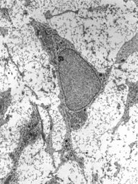
One of the main challenges in cell therapy for muscle diseases is to efficiently target the muscle. To address this issue and achieve better understanding of in vivo cell fate, we evaluated the relevance of a non-invasive cell tracking method in the Golden Retriever Muscular Dystrophy (GRMD) model, a well-recognised model of Duchenne Muscular Dystrophy (DMD). Mesoangioblasts were directly labelled with 111In-oxine, and injected through one of the femoral arteries. The scintigraphy images obtained provided the first quantitative mapping of the immediate biodistribution of mesoangioblasts in a large animal model of DMD. The results revealed that cells were trapped by the first capillary filters: the injected limb and the lung. During the days following injection, radioactivity was redistributed to the liver. In vitro studies, performed with the same cells prepared for injecting the animal, revealed prominent cell death and 111In release. In vivo, cell death resulted in 111In release into the vasculature that was taken up by the liver, resulting in a non-specific and non-cell-bound radioactive signal. Indirect labelling methods would be an attractive alternative to track cells on the mid- and long-term.
Journal Title: Scientific Reports
Year Published: 2020
Link to full text (if available)
Share on Social Media: Sign Up to like & get
recommendations!