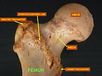
Photo from wikipedia
This longitudinal observational study investigated the relationship between changes in spinal sagittal alignment and changes in lower extremity coronal alignment. A total of 58 female volunteers who visited our institution… Click to show full abstract
This longitudinal observational study investigated the relationship between changes in spinal sagittal alignment and changes in lower extremity coronal alignment. A total of 58 female volunteers who visited our institution at least twice during the 1992 to 1997 and 2015 to 2019 periods were investigated. We reviewed whole-spine radiographs and lower extremity radiographs and measured standard spinal sagittal parameters including pelvic incidence [PI], lumbar lordosis [LL], pelvic tilt [PT], sacral slope [SS] and sagittal vertical axis [SVA], and coronal lower extremity parameters including femorotibial angle (FTA), hip–knee–ankle angle (HKA), mechanical lateral distal femoral angle (mLDFA), mechanical medial proximal tibial angle (mMPTA) and mechanical lateral distal tibial angle (mLDTA). Lumbar spondylosis and knee osteoarthritis were assessed using the Kellgren–Lawrence (KL) grading system at baseline and at final follow-up. We investigated the correlation between changes in spinal sagittal alignment and lower extremity alignment and changes in lumbar spondylosis. The mean age [standard deviation (SD)] was 48.3 (6.3) years at first visit and 70.2 (6.3) years at final follow-up. There was a correlation between changes in PI-LL and FTA (R = 0.449, P < 0.001) and between PI-LL and HKA (R = 0.412, P = 0.001). There was a correlation between changes in lumbar spondylosis at L3/4 (R = 0.383, P = 0.004) and L4/5 (R = 0.333, P = 0.012) and the knee joints. Changes in lumbar spondylosis at L3/4 and L4/5 were related to changes in KOA. Successful management of ASD must include evaluation of the state of lower extremity alignment, not only in the sagittal phase, but also the coronal phase.
Journal Title: Scientific Reports
Year Published: 2020
Link to full text (if available)
Share on Social Media: Sign Up to like & get
recommendations!