Photo from wikipedia
Ternary I–III–VI nanocrystals (NCs) without toxic elements, such as Cu–In–S, are promising candidates for biolabeling and bioimaging due to their favorable biocompatibility. At present, it remains a tremendous challenge to… Click to show full abstract
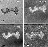
Ternary I–III–VI nanocrystals (NCs) without toxic elements, such as Cu–In–S, are promising candidates for biolabeling and bioimaging due to their favorable biocompatibility. At present, it remains a tremendous challenge to directly synthesize high-quality Cu–In–S/ZnS core/shell NCs in aqueous solution at atmospheric pressure. Herein, a simple and reliable strategy that combines the hot-injection method and successive ion layer adsorption and reaction has been proposed to realize aqueous synthesis of fluorescent Cu–In–S/ZnS core/shell NCs. The growth of ZnS shell around Cu–In–S NCs not only improved the fluorescence quantum yield by several hundred times and to the value of 18%, but also effectively enhanced their fluorescence stability by protecting Cu+ in Cu–In–S core from being oxidized to Cu2+. The prepared Cu–In–S/ZnS core/shell NCs with bright and stable fluorescence were conjugated with wheat germ agglutinin and further applied to cancer cell imaging. This strategy here should open new avenues for aqueous synthesis of high-quality nanomaterials with the core/shell structure for broad biomedical applications.
Journal Title: RSC Advances
Year Published: 2017
Link to full text (if available)
Share on Social Media: Sign Up to like & get
recommendations!