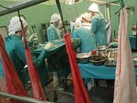
Photo from wikipedia
OBJECTIVES The use of bioartificial lungs may represent a breakthrough for the treatment of end-stage lung disease. The present study aimed to evaluate the feasibility of transplanting bioengineered lungs created… Click to show full abstract
OBJECTIVES The use of bioartificial lungs may represent a breakthrough for the treatment of end-stage lung disease. The present study aimed to evaluate the feasibility of transplanting bioengineered lungs created from autologous cells. METHODS Porcine decellularized lung scaffolds were seeded with porcine recipient-derived airway and vascular cells. The porcine recipient-derived cells were collected from lung tissue obtained by pulmonary wedge resection. Following culture of autologous cells in the scaffolds, the resulting grafts were unilaterally transplanted into porcine recipients (n=3). Allograft left unilateral lung transplantation was performed in the control group (n=3). Left unilateral transplantation of decellularized grafts was also performed in a separate control group (n=2). In vivo functions were assessed for 2 hours after transplantation. RESULTS Histological evaluation and immunostaining showed the presence of airway and vascular cells in the bioengineered lungs. No animals survived in the decellularized transplant group, whereas all animals survived in the bioengineered transplant and allotransplant groups. However, bioengineered lung grafts showed marked bullous changes. The oxygen exchange was comparable between the bioengineered lung graft transplant and allograft transplant groups. However, the carbon dioxide gas exchange of the bioengineered lung graft transplant group was significantly lower than that of the allograft transplant group at 2 hours after transplantation (4.10±0.87 mmHg vs 24.7±10.1 mmHg, P=0.02). CONCLUSIONS Transplantation of bioartificial lung grafts created from autologous cells was feasible in the super-acute phase. However, bullous changes and poor carbon dioxide gas exchange remain limitations of this method.
Journal Title: Seminars in thoracic and cardiovascular surgery
Year Published: 2020
Link to full text (if available)
Share on Social Media: Sign Up to like & get
recommendations!