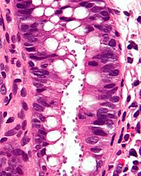
Photo from wikipedia
Immunosuppressed patients are susceptible to infections by opportunistic agents such as Leishmania that could cause visceral leishmaniasis with gastrointestinal involvement in up to 10% of cases. We report a 41-year-old… Click to show full abstract
Immunosuppressed patients are susceptible to infections by opportunistic agents such as Leishmania that could cause visceral leishmaniasis with gastrointestinal involvement in up to 10% of cases. We report a 41-year-old man with human immunodeficiency virus (HIV) infection stage C3 with CD4 lymphocytes 81/mm3, 4,070 leukocytes (47.2% lymphocytes, 0.0% eosinophils, rest of differential normal) treated with antiretroviral therapy (dolutegravir/abacavir/lamivudine) with good adherence. He also reported mesangiocapillary glomerulonephritis type-1, hepatocutaneous porphyria, and a 7-year history of recurrent visceral leishmaniasis treated with liposomal amphotericin B as secondary prophylaxis. Esophagogastroduodenoscopy and colonoscopy indicated for chronic diarrhea and anemia performed 5 years ago displayed antral erythema, mild nodular appearance in the duodenal mucosa, and normal colonic mucosa. Gastric, duodenal, and colonic biopsies revealed Leishmania spp despite treatment with liposomal amphotericin B. A video capsule endoscopy (VCE) was now indicated for persistent diarrhea. Enteropathy with atrophic and patchy, marked edema of the villus, and whitish nodularity with a “river bedrock” appearance (▶Fig. 1–3) in the duodenum and jejunum were identified (▶Video 1). Further gastric and duodenal biopsies showed an accumulation of macrophages in the lamina propria of the mucosa with intracytoplasmatic Leishmania spp (▶Fig. 4). Treatment withmeglumine antimoniate was initiated owing to previous failure with liposomal amphotericin B, without response. Some cases of visceral leishmaniasis showing non-specific findings (atrophy, edema, and whitish nodular mucosa) on esophagogastroduodenoscopy have been reported [1, 2], with the mucosa appearing normal in up of 45% of cases [3, 4]. There is only one case reporting E-Videos
Journal Title: Endoscopy
Year Published: 2021
Link to full text (if available)
Share on Social Media: Sign Up to like & get
recommendations!