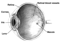
Photo from wikipedia
BACKGROUND The aim of this study was to evaluate the thickness of the outer retinal layer (ORL) together with macular thickness and changes in the retinal nerve fiber layer (RNFL)… Click to show full abstract
BACKGROUND The aim of this study was to evaluate the thickness of the outer retinal layer (ORL) together with macular thickness and changes in the retinal nerve fiber layer (RNFL) in patients with schizophrenia in comparison with healthy controls. METHODS This study included 114 eyes of 57 patients diagnosed with schizophrenia and 114 eyes of 57 healthy controls. Central foveal thickness (CFT), central macular thickness (CMT), and ORL thickness were measured in both groups via the images obtained by spectral-domain optical coherence tomography (SD-OCT). RNFL was also assessed in four quadrants (inferior, superior, temporal, nasal). CMT measurements were presented as the average thickness of the macula in the central 1 mm area on the Early Treatment Diabetic Retinopathy Study (ETDRS) grid. The ORL thickness was defined as the distance between the external limiting membrane and retinal pigment epithelium at the center of the foveal pit. RESULTS The mean age of 57 patients was 37 ± 10 years, of whom 34 (60%) were male and 23 (40%) female. No statistically significant difference was found between groups in terms of age and gender (p = 0.8 for age, p = 0.9 for gender). There was no statistically significant difference in the mean CMT between the two groups (p = 0.1). The mean ORL thickness in the two groups was 99.8 ± 8.3 and 103.7 ± 6.2, respectively, and was significantly decreased in the schizophrenia group (p = 0.005). RNFL analysis demonstrated significant thinning in the inferior and superior quadrants compared to healthy controls (p < 0.001 and p = 0.017, respectively). CONCLUSIONS SD-OCT findings - especially ORL and RNFL thickness - may be related to the neurodegenerational changes in schizophrenia.
Journal Title: Klinische Monatsblatter fur Augenheilkunde
Year Published: 2022
Link to full text (if available)
Share on Social Media: Sign Up to like & get
recommendations!