Photo from wikipedia
The efficacy and safety of endoscopic submucosal dissection (ESD) for treating cervical esophageal carcinoma (CEC) are well recognized [1, 2]. However, performing ESD in the cervical esophagus remains challenging due… Click to show full abstract
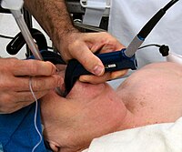
The efficacy and safety of endoscopic submucosal dissection (ESD) for treating cervical esophageal carcinoma (CEC) are well recognized [1, 2]. However, performing ESD in the cervical esophagus remains challenging due to upper sphincter contraction and endotracheal intubation balloon compression. Herein, a deeper endotracheal intubation was applied to tackle this technical difficulty. A 76-year-old man diagnosed with CEC was referred to our hospital for endoscopic resection (▶Fig. 1). An ultrafine endoscope (GIF-XP260NS; Olympus Corp., Tokyo, Japan) was used to conduct endotracheal intubation. The endotracheal intubation tube was inserted above the tracheal carina, 30 cm from the incisor teeth, and the balloon was located 26–30cm from the incisors after inflation (▶Fig. 2, ▶Video 1). In a procedure lasting 32 minutes, the lesion underwent en bloc resection with standard ESD (▶Fig. 3). The resected specimen was confirmed as squamous cell carcinoma with invasion to the lamina propria mucosa but without lymphovascular invasion, and with free margins. Traditionally, endotracheal intubation is adopted during cervical esophageal ESD to avoid aspiration, but the balloon compression position is 17–22cm from the incisors, where the cervical and upper thoracic esophagus are located. In our case, two measures were taken to relieve the compression from the balloon: 1. the ultrafine endoscope was used to visualize the accurate position of the tube and avoid the blindness caused by laryngoscope intubation in traditional endotracheal intubation; 2. the tube was inserted above the tracheal carina to relieve the compression of the balloon on the cervical esophagus. The improvement of vision and the expansion of working space allowed avoidance of intraoperative bleeding and muscularis propria injury. This is the first report of endotracheal intubation using an ultrafine endoscope, and ESD conditions were improved by deepening the position of intubation.
Journal Title: Endoscopy
Year Published: 2023
Link to full text (if available)
Share on Social Media: Sign Up to like & get
recommendations!