Photo from wikipedia
BACKGROUND Cerebral venous thrombosis (CVT) is a rare disease, and with poor prognosis. Computed tomography (CT) and magnetic resonance imaging (MRI) are the most commonly used image modalities for patients… Click to show full abstract
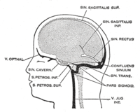
BACKGROUND Cerebral venous thrombosis (CVT) is a rare disease, and with poor prognosis. Computed tomography (CT) and magnetic resonance imaging (MRI) are the most commonly used image modalities for patients with non-specific neurologic symptoms. We present here a meta-analysis to assess the accuracy of CT and MRI in the differential diagnosis of CVT and cerebral venous sinus thrombosis (CVST). MATERIALS AND METHODS A comprehensive search of the PubMed, EMBASE, Web of Science, Cochrane Database and Chinese Biomedical (CBM) databases was conducted prior to March 20, 2017. In this report, we assess the methodological quality of each article individually and perform a meta-analysis to obtain the summary of the diagnostic accuracy of CT and MRI in correctly identifying CVT and CVST. RESULTS Twenty-four eligible articles comprising 48 studies (4,595 cases) were included. The pooled sensitivity for CT-CVT/CT-CVST groups is 0.79 (95% confidence interval [CI]: 0.76, 0.82)/0.81(95% CI: 0.78, 0.84), and pooled specificity is 0.90 (95% CI: 0.89, 0.91)/0.89 (0.88, 0.91), with an area under the curve (AUC) for the summary receiver operating characteristic (SROC) of 0.9314/0.9161, respectively. No significant heterogeneity and publication bias was observed across each study. For MRI-CVT/MRI-CVST, the pooled sensitivity is 0.82 (95% CI: 0.78, 0.85)/0.80 (95% CI: 0.76, 0.83), and pooled specificity is 0.92 (95% CI: 0.91, 0.94)/0.91(0.89, 0.92), with an AUC for the SROC of 0.9221/0.9273, respectively. CONCLUSION This meta-analysis indicates that both CT and MRI have a high level of diagnostic accuracy in the differential diagnosis of CVT and CVST, independent of stage, target for analysis or analysis methods. They could be chosen as alternative sub-optimal gold standards for diagnosing CVT and CVST, especially in emergency.
Journal Title: Thrombosis and haemostasis
Year Published: 2018
Link to full text (if available)
Share on Social Media: Sign Up to like & get
recommendations!