Photo from wikipedia
The usefulness of endoscopic ultrasound (EUS) as a diagnostic tool is well known; however, its therapeutic implications are upcoming. There are data for the use of cyanoacrylate glue (CYA) as… Click to show full abstract
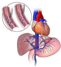
The usefulness of endoscopic ultrasound (EUS) as a diagnostic tool is well known; however, its therapeutic implications are upcoming. There are data for the use of cyanoacrylate glue (CYA) as a temporary measure to control bleeding in gastric varices, followed either by a transjugular intrahepatic portosystemic shunt (TIPS) procedure or by balloon-occluded retrograde transvenous obliteration (BRTO). A 44-year-old Asian man with hepatitis B virus cirrhosis complicated by hepatocellular cancer causing portal vein thrombosis presented with melena and bright red blood per rectum with associated dizziness, hemodynamic instability, and an initial hemoglobin of 6.5 g/dL. He had a history of bleeding esophageal varices that had been banded in the past, with prior EGD showing non bleeding gastric varices. An emergent repeat esophagogastroduodenoscopy showed no residual esophageal varices and the stomach was filled with blood clots and fresh blood that were preventing identification of the bleeding source, but he had known prior gastric varices making that to be the most likely source of bleeding. An emergent EUS showed multiple gastric varices with active blood flow (▶Fig. 1). CYA was unavailable and, in view of the patient’s hemodynamic instability, a decision was made to emergently inject 3% sodium tetradecyl sulfate (STS), a sclerosing agent, under EUS guidance. Doppler ultrasound confirmed a significant decrease in the blood flow to the gastric varices. Following this an interventional radiology opinion was sought, but the patient was deemed a poor candidate for TIPS and BRTO because of his portal vein thrombosis and advanced cirrhosis. The following day, on repeat EUS, two deep gastric varices were therefore identified and injected with two tornado 4-mm× 30-mm coils, followed by a further 3mL E-Videos
Journal Title: Endoscopy
Year Published: 2018
Link to full text (if available)
Share on Social Media: Sign Up to like & get
recommendations!