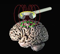
Photo from wikipedia
Significance Transcranial magnetic stimulation (TMS) holds promise as a tool for noninvasively facilitating plastic changes in cortical networks. However, highly resolved visualization of its modulatory effects remains elusive because current… Click to show full abstract
Significance Transcranial magnetic stimulation (TMS) holds promise as a tool for noninvasively facilitating plastic changes in cortical networks. However, highly resolved visualization of its modulatory effects remains elusive because current neuroimaging techniques applicable in humans are limited in spatiotemporal resolution. Here we used an imaging approach with voltage-sensitive dye and tracked, at submillimeter range, TMS-induced plastic changes across cat primary visual cortex. We show that high-frequency 10-Hz TMS induces a state where visual cortical maps are transiently “destabilized.” In turn, the cortex was sensitized to a bias in input—here imposed by prolonged exposure to a single visual orientation—and primed to relearn connectivity patterns. These findings implicate an early post-TMS time window for promising therapeutic interventions through TMS. Transcranial magnetic stimulation (TMS) has become a popular clinical method to modify cortical processing. The events underlying TMS-induced functional changes remain, however, largely unknown because current noninvasive recording methods lack spatiotemporal resolution or are incompatible with the strong TMS-associated electrical field. In particular, an answer to the question of how the relatively unspecific nature of TMS stimulation leads to specific neuronal reorganization, as well as a detailed picture of TMS-triggered reorganization of functional brain modules, is missing. Here we used real-time optical imaging in an animal experimental setting to track, at submillimeter range, TMS-induced functional changes in visual feature maps over several square millimeters of the brain’s surface. We show that high-frequency TMS creates a transient cortical state with increased excitability and increased response variability, which opens a time window for enhanced plasticity. Visual stimulation (i.e., 30 min of passive exposure) with a single orientation applied during this TMS-induced permissive period led to enlarged imprinting of the chosen orientation on the visual map across visual cortex. This reorganization was stable for hours and was characterized by a systematic shift in orientation preference toward the trained orientation. Thus, TMS can noninvasively trigger a targeted large-scale remodeling of fundamentally mature functional architecture in early sensory cortex.
Journal Title: Proceedings of the National Academy of Sciences of the United States of America
Year Published: 2018
Link to full text (if available)
Share on Social Media: Sign Up to like & get
recommendations!