Photo from wikipedia
Abstract Introduction: The anatomy of the accessory nerve has been well described but continued new clinical and anatomical findings exemplify our lack of a full understanding of the course of… Click to show full abstract
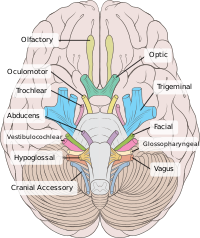
Abstract Introduction: The anatomy of the accessory nerve has been well described but continued new clinical and anatomical findings exemplify our lack of a full understanding of the course of this nerve. Therefore, this study aimed to expand on our knowledge of the course of the 11th cranial nerve via anatomical dissections. Methods: Fifty-six cadavers (112 sides) underwent dissection of the accessory nerve from its cranial and spinal origins to its emergence into the posterior cervical triangle. Immunohistochemistry was performed when appropriate. Results: Our findings included two cases (1.8%) where the nerve was duplicated, one intracranially and one extracranially. One accessory nerve (0.9%) was found to enter its own dural compartment within the jugular foramen. The majority of sides (80%) were found to have a cranial root of the accessory nerve. Thirty-one sides (28%) had connections to cervical dorsal roots medially and three sides (2.7%) laterally. Medial connections were most common with the C1 nerve. Medial components of these dorsal root connections were all sensory in nature. However, lateral components were motor on two sides (1.8%). Nerves traveled anterior to the internal jugular vein on 88% of sides. One (0.9%) left side nerve joined an interneural anastomosis between the dorsal rootlets. Macroganglia were found on the spinal part of the intracranial nerve on 13% of sides. The lesser occipital nerve arose directly from the accessory nerve on two sides (1.8%) and communicated with the accessory nerve on 5.4% of sides. One side (0.9%) was found to communicate with the facial nerve with both nerves innervating the sternocleidomastoid muscle. Conclusions: Additional anatomical knowledge of the variants of the accessory nerve may benefit patient care when this nerve is pathologically involved.
Journal Title: British Journal of Neurosurgery
Year Published: 2017
Link to full text (if available)
Share on Social Media: Sign Up to like & get
recommendations!