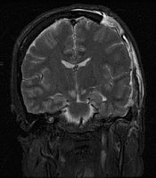
Photo from wikipedia
OBJECTIVE To determine the border of glial tumors by diffusion weighted imaging (DWI), apparent diffusion co-efficient (ADC), magnetic resonance spectroscopy (MRS) and perfusion brain MRI. PATIENTS AND METHODS Ten patients… Click to show full abstract
OBJECTIVE To determine the border of glial tumors by diffusion weighted imaging (DWI), apparent diffusion co-efficient (ADC), magnetic resonance spectroscopy (MRS) and perfusion brain MRI. PATIENTS AND METHODS Ten patients with brain gliomas were enrolled [mean age: 35.3 ± 13.2, range: 20-62]. Conventional MRI was performed for all patients. Besides, tumor mapping based on Choline (Cho)/Creatine (Cr) color map in MRS, perfusion and diffusion color maps, were gathered. Different tumoral and peritumoral regions [normal tissue, reactive edema, infiltrative edema, and tumor core] were defined. MRI criteria were evaluated in areas targeted for biopsy and histopathologic evaluation was determined. RESULTS Tumor cell positive samples [one necrosis, 26 infiltrative and nine tumor cores] composed 36 (75%) of the 48 samples. Seven (19.4%) of the positive samples were interpreted as not tumor on MRI. Five were identified as reactive edema and two as normal tissue] [kappa: .67, p-value < .001]. Mean of ADC, median of N-acetylaspartate (NAA) and NAA/Cho were statistically different between positive and negative samples (p = .02 and p < .001, respectively). Mean ADC and median Cho/NAA were statistically different in missed tumor containing tissue presented as reactive edema compared to normal and correctly diagnosed reactive edema samples together (p-values < .05). CONCLUSIONS Multimodal MRI could define infiltrated borders of brain gliomas.
Journal Title: British journal of neurosurgery
Year Published: 2020
Link to full text (if available)
Share on Social Media: Sign Up to like & get
recommendations!