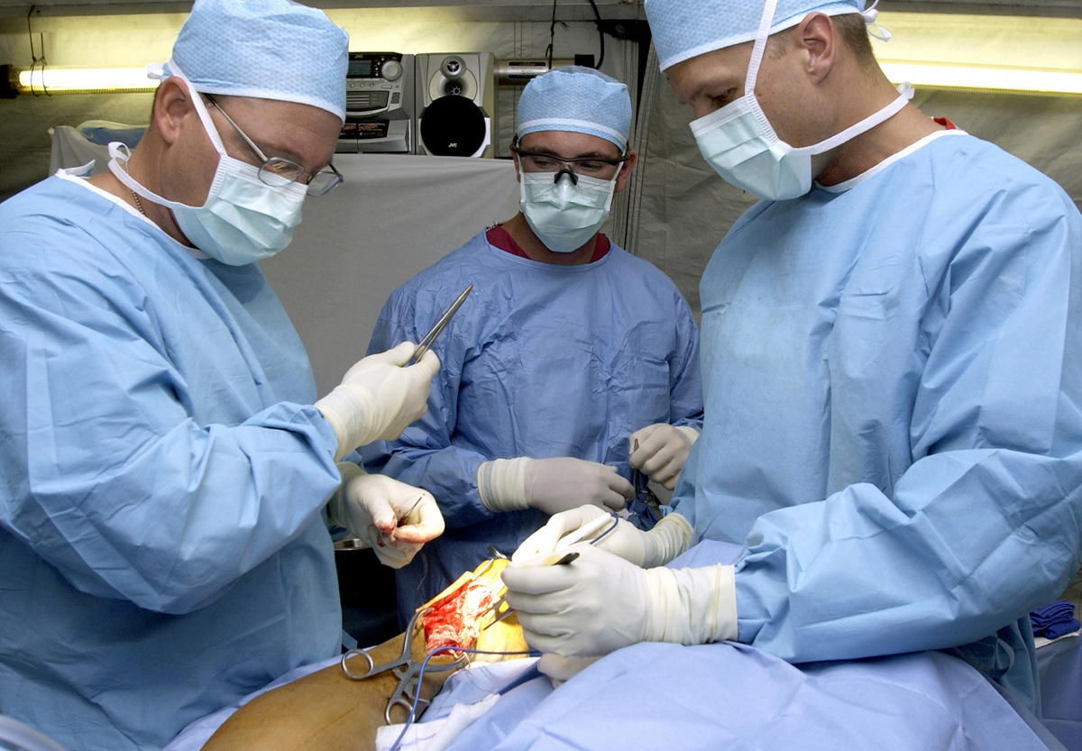
Photo from academic.microsoft.com
ABSTRACT Calcaneal fractures are amongst the most complex injuries known to man. Their intricate anatomy and extensive damage after trauma make them difficult to understand and treat. Most surgeons specialized… Click to show full abstract
ABSTRACT Calcaneal fractures are amongst the most complex injuries known to man. Their intricate anatomy and extensive damage after trauma make them difficult to understand and treat. Most surgeons specialized in foot and ankle trauma agree that in most patients surgical managements yields the best result. Functional outcome is largely dependent on preventing complications and restoring anatomy. Reconstruction of height and suntalar joint congruency for example are both associated with improved outcome. Over the years insight in the complex (patho-)anatomy has increased. First by conventional radiographs, later with computed tomography. Recently 3D scans and prints have been added to this armamentarium. The study in the current issue of the Journal of Investigative Surgery explores the use of 3D printed calcaneal fractures and the effect on restoring anatomy and functional outcome. An invited short commentary was provided.
Journal Title: Journal of Investigative Surgery
Year Published: 2018
Link to full text (if available)
Share on Social Media: Sign Up to like & get
recommendations!