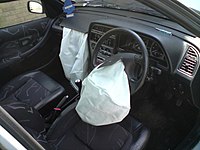
Photo from wikipedia
Abstract Objective Poor seat belt fit can result in submarining behavior and injuries to the lower extremity and abdomen. While previous studies have explored seat belt fit relative to skeletal… Click to show full abstract
Abstract Objective Poor seat belt fit can result in submarining behavior and injuries to the lower extremity and abdomen. While previous studies have explored seat belt fit relative to skeletal landmarks using palpation, medical imaging remains the gold standard for visualizing and locating skeletal landmarks and soft tissues. The goal of this study was to create a method to image automotive postures and seat belt fit from the pelvis to the clavicle using an Upright Open MRI. Methods The posture and belt fit of 10 volunteers (5M, 5F) were measured in an Acura TLX in each subject's preferred driving posture and a standard reclined posture, and then reproduced in a custom non-ferromagnetic seat replica in the MR scanner with an MRI-visible seat belt. The MRI sequence and coil placement were designed to yield clear visualization of bone, soft tissue borders, and the seat belt markers in separate scans of the pelvis, lumbar, thoracolumbar, and thoracic regions. A process was developed to precisely register the scans, and methods for digitizing spinal and pelvic landmarks were established to quantify belt fit. Conclusions This method creates opportunities to study variation in seat belt fit in different automotive postures, for occupants of different sexes, ages, BMIs, anthropometries, and for pregnant occupants.
Journal Title: Traffic Injury Prevention
Year Published: 2022
Link to full text (if available)
Share on Social Media: Sign Up to like & get
recommendations!