Photo from wikipedia
Abstract Purpose We investigated corneal endothelial morphology and corneal densitometry in smokers and compared our results with findings observed in non-smokers. Materials and Methods This cross-sectional observational study included 100… Click to show full abstract
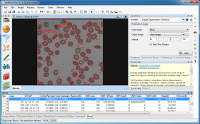
Abstract Purpose We investigated corneal endothelial morphology and corneal densitometry in smokers and compared our results with findings observed in non-smokers. Materials and Methods This cross-sectional observational study included 100 participants (50 smokers, 50 non-smokers) aged 18–80 years in whom corneal endothelial morphology was analysed using a non-contact Tomey EM-4000 specular microscope (Tomey Corporation, Japan). The Pentacam HR system was used to measure corneal densitometry spatially in three concentric zones (from the centre to the periphery) and at three different corneal depths (from the anterior to the posterior aspects). Endothelial morphology findings and corneal densitometry values were recorded in all participants, and these results were compared between smokers and non-smokers. Results Endothelial morphology and corneal densitometry analysis showed significantly lower endothelial cell counts (Num) in smokers (228 cells/mm2 vs. 246 cells/mm2, p = 0.02) in addition to increased maximum cell area (Max) values (986.5 µm2 vs. 935 µm2, p = 0.04). We observed no statistically significant intergroup difference in corneal densitometry values (p > 0.05 for each zone); however, we observed a moderately positive correlation between densitometry values in the 6–10 mm concentric zone and between the all total corneal zones and number of pack-years in smokers. Conclusions Our study highlights that among the morphometric corneal endothelial variables analysed in this study, only the Num value was significantly correlated with smoking. We observed no statistically significant intergroup difference in corneal densitometry values in this study; however, a positive correlation was observed between the number of pack-years and corneal densitometry findings. Therefore, as the pack-years increase, the increase in corneal densitometry values may indicate a decrease in corneal clarity, considering the possible contribution of secondary factors such as age.
Journal Title: Cutaneous and Ocular Toxicology
Year Published: 2021
Link to full text (if available)
Share on Social Media: Sign Up to like & get
recommendations!