Photo from wikipedia
Tetralogy of Fallot (TOF) occurs in 4 of every 10,000 live births and is the most common form of cyanotic congenital heart disease. Patients with repaired TOF (rTOF) require long-term… Click to show full abstract
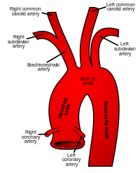
Tetralogy of Fallot (TOF) occurs in 4 of every 10,000 live births and is the most common form of cyanotic congenital heart disease. Patients with repaired TOF (rTOF) require long-term and frequent monitoring for many complications that may arise. The hemodynamic alterations that contribute to the quality of life and outcomes for these patients are understudied and poorly understood. The objective of this study was to use 4D Flow MRI to assess flow hemodynamics in patients with rTOF to better identify and predict altered hemodynamic patterns to assist with future interventions. We hypothesized, patients with rTOF will have abnormal left-sided flow hemodynamics compared to healthy controls resulting in poorer hemodynamic patterns even after post-repair. A total of 20 rToF patients (age = 34.5±11.2, female = 5) and 20 healthy controls (age = 37.0±12.1, female = 6) were enrolled in this study and underwent standard cardiac MRI followed by 4D Flow MRI acquisition. Figure 1 demonstrates the workflow of the analysis that was performed using cvi42 v5.11 (Circle Cardiovascular Imaging Inc., Calgary, Canada). The Aorta and LV were segmented, flow visualization and quantitative flow analysis were performed by placing analysis planes perpendicular to the flow of interest as shown in Figure 1. Total volume (TV), Wall Shear Stress Axial (WSSax), circumferential (WSScirc) and energy loss (EL) were calculated. Statistics were analyzed using IBM SPSS Statistics, version 27. An independent-samples t-test was used to compare parameters and identify significant differences between controls and patients. A P-value <0.05 was considered significant. In comparison to controls, TV of the STJ (66.89±17.33 vs. 82.28±18.77, p=0.011), Aao (56.05±10.71 vs. 73.04±19.66, p=0.002), and 1st Aortic Arch (AAr) (56.88±12.97 vs. 69.52±18.65, p=0.017) were lower in rTOF patients. In addition, patients with rTOF had higher average WSSax in the LVOT (0.13±0.05 vs. 0.10±0.03, p=0.049), STJ (0.10±0.02 vs. 0.07±0.02, p=0.001), and Aao (0.10±0.03 vs. 0.08±0.02, p<0.000) compared to controls. Moreover, average WSScirc in the LVOT (0.07±0.02 vs. 0.05±0.01, p=0.010), STJ (0.07±0.02 vs. 0.05±0.01, p=0.006), Aao (0.07±0.02 vs. 0.05±0.01, p=0.004), and 1st AAr (0.06±0.02 vs. 0.05±0.01, p=0.017) were higher in patients compared to controls. Lastly, EL in the Aao was lower in patients compared to controls (1.87±0.83 vs. 2.47±1.03, p=0.049). Significant results are demonstrated in Table 1, red illustrating lower values in patients compared to controls and green illustrating higher values. This study unveiled abnormal left-sided blood flow in rToF patients with reduced TV and increased WSSax, average WSScirc and EL. These new hemodynamic insights obtained from 4D flow MRI may help to inform future individualized decision-making for patients with rTOF. Type of funding sources: Public Institution(s). Main funding source(s): University of Calgary
Journal Title: European Heart Journal
Year Published: 2021
Link to full text (if available)
Share on Social Media: Sign Up to like & get
recommendations!