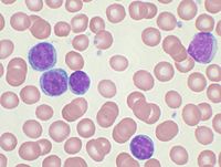
Photo from wikipedia
A 71–year–old woman with chronic lymphocytic leukaemia (CLL) presented to our Cardiology clinic complaining of recent–onset chest pain and dysphonia. Physical examination and ECG were normal. A blood test revealed… Click to show full abstract
A 71–year–old woman with chronic lymphocytic leukaemia (CLL) presented to our Cardiology clinic complaining of recent–onset chest pain and dysphonia. Physical examination and ECG were normal. A blood test revealed high levels of high–sensitivity troponin T (373 ng/L) and of C–reactive protein (7 mg/dL); blood counts and differential were normal. Transthoracic echocardiography showed a hyperechogenic appearance of the interatrial septum, the interventricular septum and the right ventricular free wall, as well as a mild circumferential pericardial effusion. Cardiac magnetic resonance imaging revealed multiple intracardiac masses involving both ventricles and the interatrial septum, with evidence of oedema and fibrosis. A chest computed tomography showed the presence of multiple cervical, pulmonary, and mediastinal masses, one of which was biopsied through video–thoracoscopy. At the histological examination, the mass mainly consisted of large pleomorphic peripheral CD10–, CD20+/–, CD79a+, PAX5+, BCL2+, BCL6–, c–MYC+ B cells, with high proliferative activity (Ki–67/MIB1+ 80%). A diagnosis of CLL transformation into diffuse large B–cell lymphoma (DLBCL), also known as Richter’s syndrome (RS), was established (Figure). Despite indication for specific haematological chemotherapy, the patient rapidly deteriorated and died few days after the diagnosis. RS is a rare evolution of CLL and of small lymphocytic lymphoma, affecting 2–8% of such patients (transformation <1%/ year), yet is burdened with very poor prognosis (median overall survival 8–16 months). Extra–nodal RS is uncommon, and cardiac involvement has been reported in only 3 cases in the literature (in none with the panel of techniques here used). Despite its rarity, CLL patients with rapidly worsening cardiac symptoms should be suspected of RS transformation involving the heart. The main differential diagnosis of cardiac RS is primary cardiac lymphoma. The characterization of cardiac masses relies on multimodality imaging, but histologic analysis is required to reach a definite diagnosis, which is essential to guide subsequent therapeutic choices.
Journal Title: European heart journal
Year Published: 2023
Link to full text (if available)
Share on Social Media: Sign Up to like & get
recommendations!