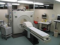
Photo from wikipedia
Even though nuclear cardiology is a non-invasive imaging method having been in clinical use for several decades, the last few years have witnessed dramatic advances regarding technical aspects and the… Click to show full abstract
Even though nuclear cardiology is a non-invasive imaging method having been in clinical use for several decades, the last few years have witnessed dramatic advances regarding technical aspects and the range of clinical application. While the main value of single photon emission computed tomography (SPECT) and positron emission tomography (PET) has for a long time been the evaluation of patients with suspected or known coronary artery disease, nowadays radionuclide imaging has gained importance in several other clinical fields of cardiology such as, assessment of infection sources (e.g. endocarditis), evaluation of infiltrative diseases (e.g. amyloidosis), and inflammation imaging (e.g. sarcoidosis). Hence, by publishing an update on nuclear cardiology 9 years after the first edition of this handbook, Drs Sabharwal, Arumugam, and Kelion provide an excellent overview for both physicians and technologists in training as well as for those with experience in the field of radionuclide imaging. This handbook on Nuclear Cardiology includes 12 chapters and is illustrated with several figures and diagrams facilitating the understanding of the topics covered. The chapters have been arranged thoughtfully. The first four chapters cover basics of nuclear cardiology, ranging from historical development and radiation physics to camera design and image reconstruction. In the following chapters radionuclide ventriculography and SPECT myocardial perfusion imaging including stress testing, different radiopharmaceuticals, and image interpretation are discussed. Chapter 11 deals with other nuclear investigations such as innervation imaging and infiltrative diseases. Particularly, as scintigraphy has proven to be extremely sensitive and specific for the diagnosis of amyloid transthyretinrelated (ATTR) amyloidosis, it is establishing itself as a simple non-invasive method of distinguishing ATTR from amyloid light chain amyloidosis. Finally, the last chapter is devoted to very important aspects on PET imaging including myocardial perfusion imaging, blood flow quantification, and viability assessment as well as on the use of F-FDG PET in inflammation imaging for cardiac sarcoidosis, and infection imaging for device and prosthetic valve infection, respectively. In conclusion, this handbook on nuclear cardiology gives a comprehensive overview on all aspects of modern cardiac imaging using radionuclide imaging. It will benefit students, trainees, technologists, and clinicians interested in the field of nuclear cardiology.
Journal Title: European heart journal
Year Published: 2018
Link to full text (if available)
Share on Social Media: Sign Up to like & get
recommendations!