Photo from wikipedia
Atrial myocardial infarction may be a risk for atrial fibrillation. However, the electrophysiological and structural characteristics of the infarcted atrial myocardium are not well known. This study aimed to analyse… Click to show full abstract
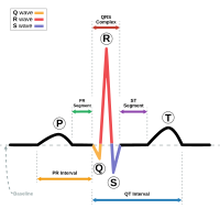
Atrial myocardial infarction may be a risk for atrial fibrillation. However, the electrophysiological and structural characteristics of the infarcted atrial myocardium are not well known. This study aimed to analyse the changes in local atrial electrograms and myocardial structure in an experimental model of isolated atrial myocardial infarction. Five anesthetized, open-chest pigs were submitted to 4 hours of acute atrial myocardial ischemia induced by direct surgical clamping of atrial coronary branches originating from the right coronary artery. In all cases, we recorded simultaneously the 12-lead surface electrocardiogram (ECG) and the epicardial mapping of local atrial electrograms (17 x 12.5 mm patches containing 128 electrodes, with 1 mm inter-electrode distance) in a region close to the occluded branches and in control non-treated atria. The changes in local atrial QRS-ST segment and the amplitude of the P-wave of the ECG were sequentially analysed (Figure). The hearts were removed and processed for anatomopathological examination. Selective atrial coronary branch occlusion induced a patchy atrial myocardial necrosis with an irregular and abrupt border zone (circled areas in the Figure). During the first 15 min of ischemia, the local atrial electrograms showed increasing R waves, widening of QRS complex, and ST segment elevation leading to monophasic potentials (maximal ST segment at 30 min: from 0.2±0.7 mV to 1.9±1.4 mV, ANOVA p<0.01). This period was followed by a phase of transient electrical recovery characterized by disappearance of monophasic potentials, reduction of ST segment elevation, and recovery of local electrical activation. After 60 min of occlusion, monophasic potentials reappeared and the magnitude of ST segment elevation decreased progressively during the ensuing 3 hours. The spatial transition from areas with monophasic potentials to normal electrograms encompassed 1 or 2 electrodes. The surface ECG showed increased duration of the P-wave (lead II at 3h occlusion: from 73.2±4.5 ms to 88.9±15.5 ms, ANOVA p<0.05) with absence of ST segment changes. Atrial arrhythmias were not observed. Structural and electrical atrial changes Selective occlusion of atrial coronary branches induced patchy atrial myocardial necrosis with abrupt anatomical and electrical border zone. The overt QRS and ST segment changes in local atrial electrograms resembled those described in acute ventricular myocardial ischemia, and were associated with widening of the P-wave on the surface ECG. Although acute ischemic arrhythmias were not observed, the atrial structural alterations might set the substrate for re-entrant arrhythmias late after the healing over process. Supported by grants from ISCI-MINECO (FISPI17/00069), CIBERCV (CB16/11/00276), FEDER and Fundaciό “La Maratό” TV3 (20150830).
Journal Title: European Heart Journal
Year Published: 2019
Link to full text (if available)
Share on Social Media: Sign Up to like & get
recommendations!