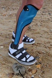
Photo from wikipedia
INTRODUCTION Knee osteoarthritis (KOA) is a primary source of long-term disability and decreased quality of life (QoL) in service members (SM) with lower limb loss (LL); however, it remains difficult… Click to show full abstract
INTRODUCTION Knee osteoarthritis (KOA) is a primary source of long-term disability and decreased quality of life (QoL) in service members (SM) with lower limb loss (LL); however, it remains difficult to preemptively identify and mitigate the progression of KOA and KOA-related symptoms. The objective of this study was to explore a comprehensive cross-sectional evaluation, at the baseline of a prospective study, for characterizing KOA in SM with traumatic LL. MATERIALS AND METHODS Thirty-eight male SM with traumatic unilateral LL (23 transtibial and 15 transfemoral), 9.5 ± 5.9 years post-injury, were cross-sectionally evaluated at initial enrollment into a prospective, longitudinal study utilizing a comprehensive evaluation to characterize knee joint health, functionality, and QoL in SM with LL. Presences of medial, lateral, and/or patellofemoral articular degeneration within the contralateral knee were identified via magnetic resonance imaging(for medically eligible SM; Kellgren-Lawrence Grade [n = 32]; and Outerbridge classification [OC; n = 22]). Tri-planar trunk and pelvic motions, knee kinetics, along with temporospatial parameters, were quantified via full-body gait evaluation and inverse dynamics. Concentrations of 26 protein biomarkers of osteochondral tissue degradation and inflammatory activity were identified via serum immunoassays. Physical function, knee symptoms, and QoL were collected via several patient reported outcome measures. RESULTS KOA was identified in 12 of 32 (37.5%; KL ≥ 1) SM with LL; however, 16 of 22 SM presented with patellofemoral degeneration (72.7%; OC ≥ 1). Service members with versus without KOA had a 26% reduction in the narrowest medial tibiofemoral joint space. Biomechanically, SM with versus without KOA walked with a 24% wider stride width and with a negative correlation between peak knee adduction moments and minimal medial tibiofemoral joint space. Physiologically, SM with versus without KOA exhibited elevated concentrations of pro-inflammatory biomarker interleukin-7 (+180%), collagen breakdown markers collagen II cleavage (+44%), and lower concentrations of hyaluronic acid (-73%) and bone resorption biomarker N-telopeptide of Type 1 Collagen (-49%). Lastly, there was a negative correlation between patient-reported contralateral knee pain severity and patient-reported functionality and QoL. CONCLUSIONS While 37.5% of SM with LL had KOA at the tibiofemoral joint (KL ≥ 1), 72.7% of SM had the presence of patellofemoral degeneration (OC ≥ 1). These findings demonstrate that the patellofemoral joint may be more susceptible to degeneration than the medial tibiofemoral compartment following traumatic LL.
Journal Title: Military medicine
Year Published: 2022
Link to full text (if available)
Share on Social Media: Sign Up to like & get
recommendations!