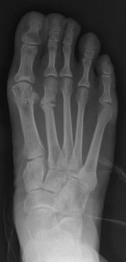
Photo from wikipedia
INTRODUCTION Military trainees are at an increased risk of stress fractures. Vitamin D availability is known to play an important role in both fracture prevention and healing. The purpose of… Click to show full abstract
INTRODUCTION Military trainees are at an increased risk of stress fractures. Vitamin D availability is known to play an important role in both fracture prevention and healing. The purpose of this investigation was to assess 25-hydroxy vitamin D (25(OH)D) levels in soldiers with confirmed lower extremity stress fractures and assess the predictors of fracture location. MATERIALS AND METHODS Following Institutional Review Board approval, military trainees at a large training base presenting to the orthopedic clinic with a radiographically verified stress fracture were identified. Demographic data and 25(OH)D levels were collected. A descriptive analysis was performed in regard to patient age, body mass index (BMI), and 25(OH)D level. Interactions between variables were assessed using one-way analysis of variance for four fracture location groups (femoral neck, femoral shaft, tibial shaft, and foot and ankle). Bivariate correlations were examined between age, BMI, and vitamin D level. RESULTS A total of 155 lower extremity stress fractures were identified in 144 males and 11 females over 30 months. The mean age was 22.7 ± 4.85 years. The majority (60.7%) of fractures were located in the femoral neck. The average 25(OH)D level was 26.8 ± 8.37 ng/mL. Overall, 26% (N = 41) of enrolled patients had normal 25(OH)D levels, 48% (N = 74) had insufficient 25(OH)D levels, and 26% (N = 40) had deficient 25(OH)D levels. Patients with femoral neck fractures and tibial shaft fractures had significantly lower BMI than patients with foot and ankle fractures (23.3 vs. 27.7, P < .001 and 24.2 vs. 27.7, P = .003, respectively). Patients with foot and ankle fractures had significantly lower 25(OH)D levels than patients with femoral shaft fractures (21.1 vs. 30.1, P = .02). There were no significant findings regarding age and fracture location. Age correlated positively (but weakly) with BMI (0.338, P < .001). There was no correlation between age and vitamin D level or BMI and vitamin D level. CONCLUSION Overall, 74% of patients in military training with lower extremity stress fractures had insufficient or deficient levels of 25(OH)D, highlighting a persistent area of concern in this population. Patients with femoral neck and tibial shaft stress fractures had significantly lower BMI than patients with foot and ankle stress fractures. This suggests that in stress fracture-prone patients, BMI may play a role in predicting fracture location.
Journal Title: Military medicine
Year Published: 2022
Link to full text (if available)
Share on Social Media: Sign Up to like & get
recommendations!