Photo from wikipedia
Recently, tumor treating fields (TTFields) were established for the treatment of newly diagnosed GBM. One of the most crucial parameters defining the treatment efficacy of TTFields is the electric field… Click to show full abstract
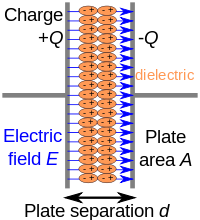
Recently, tumor treating fields (TTFields) were established for the treatment of newly diagnosed GBM. One of the most crucial parameters defining the treatment efficacy of TTFields is the electric field intensity. The dielectric properties of the normal intracranial compartments are well established, allowing the prediction of the electric field distribution. In contrast, there is no data available about the dielectric properties of brain tumor tissue. In this study we determined the dielectric properties of brain tumors by analyzing resected tissue following a fast acquisition protocol. To account for the intratumoral heterogeneity, different regions of the tumor were analyzed separately. A cohort of 84 patients with tumors of different histology and malignancy grade have been recruited (meningioma: n=26; brain metastases n=18; low grade glioma n=6; glioblastoma n=34). Tissue probes were acquired whenever possible from the vital tumor area, and perinecrotic compartment identified intraoperatively using neuronavigation, intraoperative ultrasound and fluorescence guidance. After acquisition, the tissue was measured immediately to avoid artifacts induced by temperature change, differences of fluid composition as well as post resection ischemia. A fragment was dissected from each tissue sample and was placed into a cylindrical cell with a known diameter. Two parallel electrodes were placed on both sides of the sample and the thickness of the tissue was measured using a micrometer. The impedance was recorded at frequencies 20Hz-1MHz using a software specifically developed for this study, which controls the LCR meter (Keysight Technologies, Santa Rosa, USA). The measured impedance was translated into dielectric properties of the sample (conductivity and relative permittivity) based on the parallel plate model, the recorded complex impedance and the geometry of the samples. Each tissue probe was fixed, H&E stained and histologically analyzed. We found significant differences between the conductivity of different types of tumors with meningiomas showing the lowest conductivity (mean conductivity [S/m]: 0.193; range: 0.327 - 0.113) and GBM tissue exhibiting the highest conductivity values (mean conductivity [S/m]: 0.402; range: 0.893 - 0.157). Consistently, the perinecrotic areas of tumors displayed lower conductivity values compared to the solid tumor compartments. Also, we found a significant intratumoral heterogeneity in tumors of one specific histological diagnosis. The dielectric properties of intracranial tumors appear to be depending on histological class and malignancy grade and show significant intratumoral heterogeneity. These results may allow a more precise modelling of electric field intensity distribution within the tumor.
Journal Title: Neuro-Oncology
Year Published: 2019
Link to full text (if available)
Share on Social Media: Sign Up to like & get
recommendations!