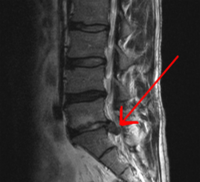
Photo from wikipedia
Thoracic disc prolapses causing cord compression can be challenging. For compressive central disc protrusions, a posterior approach is not suitable due to an unacceptable level of cord manipulation. An anterolateral… Click to show full abstract
Thoracic disc prolapses causing cord compression can be challenging. For compressive central disc protrusions, a posterior approach is not suitable due to an unacceptable level of cord manipulation. An anterolateral transthoracic approach provides direct access to the disc prolapse allowing for decompression without disturbing the spinal cord. In this video, we describe 2 cases of thoracic myelopathy from a compressive central thoracic disc prolapse. In both cases, informed consent was obtained. Despite similar radiological appearances of heavy calcification, intraoperatively significant differences can be encountered. We demonstrate different surgical strategies depending on the consistency of the disc and the adherence to the thecal sac. With adequate exposure and detachment from adjacent vertebral bodies, soft discs can be, in most instances, separated from the theca with minimal cord manipulation. On the other hand, largely calcified discs often present a significantly greater challenge and require thinning the disc capsule before removal. In cases with significant adherence to dura, in order to prevent cord injury or cerebrospinal fluid leak a thinned shell can be left, providing total detachment from adjacent vertebrae can be achieved. Postoperatively, the first patient, with a significantly calcified disc, developed a transient left leg weakness which recovered by 3-month follow-up. This video outlines the anatomical considerations and operative steps for a transthoracic approach to a central disc prolapse, whilst demonstrating that computed tomography appearances are not always indicative of potential operative difficulties.
Journal Title: Operative neurosurgery
Year Published: 2019
Link to full text (if available)
Share on Social Media: Sign Up to like & get
recommendations!