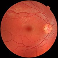
Photo from wikipedia
Purpose: To establish a multilabel-based deep learning (DL) algorithm for automatic detection and categorization of clinically significant peripheral retinal lesions using ultrawide-field fundus images. Methods: A total of 5958 ultrawide-field… Click to show full abstract
Purpose: To establish a multilabel-based deep learning (DL) algorithm for automatic detection and categorization of clinically significant peripheral retinal lesions using ultrawide-field fundus images. Methods: A total of 5958 ultrawide-field fundus images from 3740 patients were randomly split into a training set, validation set, and test set. A multilabel classifier was developed to detect rhegmatogenous retinal detachment, cystic retinal tuft, lattice degeneration, and retinal breaks. Referral decision was automatically generated based on the results of each disease class. t-distributed stochastic neighbor embedding heatmaps were used to visualize the features extracted by the neural networks. Gradient-weighted class activation mapping and guided backpropagation heatmaps were generated to investigate the image locations for decision-making by the DL models. The performance of the classifier(s) was evaluated by sensitivity, specificity, accuracy, F1 score, area under receiver operating characteristic curve (AUROC) with 95% CI, and area under the precision-recall curve. Results: In the test set, all categories achieved a sensitivity of 0.836–0.918, a specificity of 0.858–0.989, an accuracy of 0.854–0.977, an F1 score of 0.400–0.931, an AUROC of 0.9205–0.9882, and an area under the precision-recall curve of 0.6723–0.9745. The referral decisions achieved an AUROC of 0.9758 (95% CI= 0.9648–0.9869). The multilabel classifier had significantly better performance in cystic retinal tuft detection than the binary classifier (AUROC= 0.9781 vs 0.6112, P < 0.001). The model showed comparable performance with human experts. Conclusions: This new DL model of a multilabel classifier is capable of automatic, accurate, and early detection of clinically significant peripheral retinal lesions with various sample sizes. It can be applied in peripheral retinal screening in clinics.
Journal Title: Asia-Pacific Journal of Ophthalmology
Year Published: 2023
Link to full text (if available)
Share on Social Media: Sign Up to like & get
recommendations!