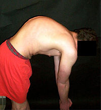
Photo from wikipedia
Objectives: To evaluate the contribution of each of the rotator cuff muscles and deltoid to fracture deformity in a 2-part proximal humerus fracture model. Our hypothesis was that superior cuff… Click to show full abstract
Objectives: To evaluate the contribution of each of the rotator cuff muscles and deltoid to fracture deformity in a 2-part proximal humerus fracture model. Our hypothesis was that superior cuff muscles would have the greatest contribution to coronal plane deformity, whereas muscles with anterior and posterior attachments would have the greatest contribution to axial and sagittal plane deformity. Methods: A medial wedge osteotomy was created in 8 cadaveric shoulder specimens. A custom shoulder testing system was used to load each rotator cuff muscle and deltoid under increasing loading conditions. Fracture displacement was measured using a Microscribe digitizing system. The primary outcome was the contribution of each muscle to varus collapse. Secondary outcomes included contributions of each muscle to apex anterior/posterior deformity and humeral head anteversion/retroversion. Results: Unbalanced loading of the supraspinatus resulted in the greatest varus deformity (34.5 ± 2.3 degrees), followed by the infraspinatus (22.3 ± 3.6 degrees) and subscapularis (21.7 ± 3.1 degree) (P < 0.05). Unbalanced loading of the subscapularis induced the greatest apex posterior (27.5 ± 4.8 degrees, P < 0.05) and retroversion (39.0 ± 5.6 degrees, P < 0.05) deformity, whereas the infraspinatus induced the greatest apex anterior (8.7 ± 3.4 degrees, P > 0.05) and anteversion (17.7 ± 5.7 degrees, P > 0.05) deformity. Conclusions: In this proximal humerus fracture model, the supraspinatus was the primary driver of varus deformity, whereas the subscapularis and infraspinatus contributed to apex posterior/retroversion and apex anterior/anteversion, respectively. The subscapularis and infraspinatus are also important secondary drivers of varus deformity. This study establishes a physiologically relevant fracture model that mimics in vivo conditions for future biomechanical testing.
Journal Title: Journal of Orthopaedic Trauma
Year Published: 2021
Link to full text (if available)
Share on Social Media: Sign Up to like & get
recommendations!