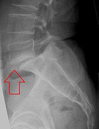
Photo from wikipedia
Study Design. A systematic review. Objective. The aim of this study was to provide an evidence-based recommendation for when and how to employ imaging studies when diagnosing back pain thought… Click to show full abstract
Study Design. A systematic review. Objective. The aim of this study was to provide an evidence-based recommendation for when and how to employ imaging studies when diagnosing back pain thought to be caused by spondylolysis in pediatric patients. Summary of Background Data. Spondylolysis is a common structural cause of back pain in pediatric patients. The radiologic methods and algorithms used to diagnose spondylolysis are inconsistent among practitioners. Methods. A literature review was performed in PubMed and Cochrane databases using the search terms “spondylolysis,” “pediatric,” “adolescent,” “juvenile,” “young,” “lumbar,” “MRI,” “bone scan,” “CT,” and “SPECT.” After inclusion criteria were applied, 13 articles pertaining to diagnostic imaging of pediatric spondylolysis were analyzed. Results. Ten papers included sensitivity calculations for comparing imaging performance. The average sensitivity of magnetic resonance imaging (MRI) with computed tomography (CT) as the standard of reference was 81.4%. When compared with single-photon emission CT (SPECT), the average sensitivity of CT was 85% and the sensitivity of MRI was 80%. Thirteen studies made a recommendation as to how best to perform diagnostic imaging of patients with clinically suspected spondylolysis. When compared with two-view plain films, bone scans had seven to nine times the effective radiation dose, while four-view plain films and CT were approximately double. Of the diagnostic methods examined, MRI was the most expensive followed by CT, bone scan, four-view plain films, and two-view plain films. Conclusion. Due to their efficacy, low cost, and low radiation exposure, we find two-view plain films to be the best initial study. With unusual presentations or refractory courses, practitioners should pursue advanced imaging. MRI should be used in early diagnosis and CT in more persistent courses. However, the lack of rigorous studies makes it difficult to formulate concrete recommendations. Level of Evidence: 3
Journal Title: SPINE
Year Published: 2017
Link to full text (if available)
Share on Social Media: Sign Up to like & get
recommendations!