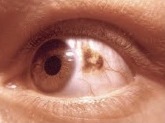
Photo from wikipedia
Purpose: To describe the imaging features of choroidal neovascularization (CNV) associated with choroidal nevus using optical coherence tomography angiography (OCT-A) imaging. Methods: Retrospective observational case series. Patients with CNV secondary… Click to show full abstract
Purpose: To describe the imaging features of choroidal neovascularization (CNV) associated with choroidal nevus using optical coherence tomography angiography (OCT-A) imaging. Methods: Retrospective observational case series. Patients with CNV secondary to choroidal nevus underwent full imaging examination including fundus photography, fluorescein angiography, indocyanine green angiography, spectral domain OCT, and OCT-A. The OCT-A features were analyzed and correlated with conventional angiography findings and spectral domain OCT. Results: There were 11 eyes from 11 patients (6 men and 5 women, mean age of 65 ± 20.4 years) included in the analysis. Fluorescein angiography and indocyanine green angiography disclosed CNV in 90% and 83%, respectively. Optical coherence tomography angiography displayed CNV network in 11 eyes (100%) and the pattern was classified as “sea-fan” in 8 (73%) and “long filamentous linear vessels” in 3 (27%) eyes. Distinct from CNV, intrinsic vasculature within the nevus was observed in six eyes (55%), corresponding to those with chronic retinal pigment epithelium changes. Conclusion: Optical coherence tomography angiography is a useful imaging technique to disclose CNV associated with choroidal nevus. Despite the presence of intraretinal or subretinal fluid and hemorrhage, OCT-A revealed the CNV in all cases, results noninferior to indocyanine green angiography. This imaging modality can be useful for analysis of long-standing nevi with related exudation.
Journal Title: Retina
Year Published: 2018
Link to full text (if available)
Share on Social Media: Sign Up to like & get
recommendations!