Photo from wikipedia
Suprachoroidal hemorrhage (SH) is an uncommon but potentially dramatic event, characterized by bleeding of the posterior ciliary vessels in the suprachoroidal space. Suprachoroidal hemorrhage may occur during ocular surgery, both… Click to show full abstract
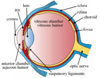
Suprachoroidal hemorrhage (SH) is an uncommon but potentially dramatic event, characterized by bleeding of the posterior ciliary vessels in the suprachoroidal space. Suprachoroidal hemorrhage may occur during ocular surgery, both intraoperatively and postoperatively. However, spontaneous SH has also been described, particularly associated with age-related macular degeneration, diabetic retinopathy, and pathologic myopia.1,2 When SH occurs spontaneously and not associated with ocular surgery, it poses a significant diagnostic challenge, which may require to rule out neoplastic disorders, including choroidal melanoma.3 Here, we describe the peculiar features on multimodal imaging of a dark-pigmented, dome-shaped choroidal lesion that spontaneously developed along the superior–temporal vascular arcade in an asymptomatic 67-year-old woman affected by pathologic myopia—32 mm axial length—(Figure 1). Of note, the lesion did not change after pressure on the globe during fundus ophthalmoscopy, and indocyanine green angiography ruled out the presence of any intrinsic vascularity or vortex vein varix; standardized echography showed medium internal reflectivity of the lesion, and optical coherence tomography illustrated its exact location, which was the suprachoroidal space. All these findings suggested the diagnosis of SH. The patient was then reassessed after three months, and the SH was almost completely reabsorbed spontaneously. Pathologic myopia has been associated with progressive elongation of the globe, increasing the risk of Fig. 1. Fundus autofluorescence, fluorescein angiography, indocyanine green angiography, and optical coherence tomography of suprachoroidal hemorrhage associated with pathologic myopia. Fundus autofluorescence (A) is relatively normal, suggesting a subretinal pigment epithelium location of the lesion. Fluorescein angiography (B) discloses physiologic retinal vascularity, with mild choroidal hypofluorescence. Indocyanine green angiography shows normal choroidal perfusion during the early phase (C), with relative hypofluorescence in the late phase in the area of a vortex ampulla (D). Optical coherence tomography at baseline (E) demonstrates a domeshaped mass in the suprachoroidal space shadowing the structures below; the neurosensory retina, the retinal pigment epithelium, and the choroid are visible above the hemorrhage. Optical coherence tomography three months after (F) shows reabsorption of most of the suprachoroidal hemorrhage with the disappearance of the dome-shaped lesion. However, a sliver of residual suprachoroidal hemorrhage is still visible (arrowheads).
Journal Title: Retina
Year Published: 2018
Link to full text (if available)
Share on Social Media: Sign Up to like & get
recommendations!