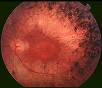
Photo from wikipedia
Supplemental Digital Content is Available in the Text. This study demonstrates important vascular and structural changes in eyes of X-linked retinoschisis patients affecting their visual outcome, with structural deterioration of… Click to show full abstract
Supplemental Digital Content is Available in the Text. This study demonstrates important vascular and structural changes in eyes of X-linked retinoschisis patients affecting their visual outcome, with structural deterioration of the photoreceptor layer, best reflecting the degenerative changes, although microvascular alteration in deep capillary plexus and foveal avascular zone shows correlation with the functional outcome. Purpose: The study aimed to evaluate the macular microvasculature of X-linked retinoschisis (XLRS) and identify correlations between vascular changes, structural changes, and functional outcome. Methods: Genetically confirmed XLRS patients and heathy control subjects underwent complete ophthalmic examination, dilated funduscopic examination, optical coherence tomography, and optical coherence tomography angiography. Schisis distribution, outer plexiform layer discontinuation, photoreceptor layer thickness, and photoreceptor outer segment length were reviewed using optical coherence tomography. Vascular flow density and foveal thickness at foveal and parafoveal area were measured using optical coherence tomography angiography. Results: A total of 17 eyes of 9 XLRS patients and 22 eyes of 11 control subjects were examined from July 2018 to August 2020. Flow density in the deep capillary plexus at foveal and parafoveal area decreased in XLRS patients compared with control subjects (P = 0.014 and 0.001, respectively), whereas foveal avascular zone area and perimeter remarkably increased (P = 0.015 and 0.001, respectively). Although outer and total retinal layers were significantly thicker in XLRS, inner retinal layer was thinner with reduced photoreceptor layer thickness and shortened photoreceptor outer segment length (P < 0.001 and P < 0.001, respectively). Foveal flow loss in deep capillary plexus, foveal avascular zone enlargement, thinner inner retina and photoreceptor layer thickness, and shortened photoreceptor outer segment length correlated with best-corrected visual acuity. Conclusion: X-linked retinoschisis eyes exhibit decreased flow density in the deep capillary plexus and variable foveal avascular zone with enlarged perimeter. Structural deterioration of the photoreceptor best reflects the degenerative changes, whereas microvascular alteration shows considerable correlation with functional outcome in XLRS.
Journal Title: Retina
Year Published: 2022
Link to full text (if available)
Share on Social Media: Sign Up to like & get
recommendations!