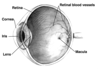
Photo from wikipedia
This prospective, cross-sectional, multicenter study analyzed choroidal vascular density obtained from swept-source optical coherence tomography en-face images of patients with diabetes, those with and without diabetic retinopathy, and healthy age-matched… Click to show full abstract
This prospective, cross-sectional, multicenter study analyzed choroidal vascular density obtained from swept-source optical coherence tomography en-face images of patients with diabetes, those with and without diabetic retinopathy, and healthy age-matched volunteers. The no diabetic retinopathy group had significantly higher choroidal vascular density values, and the proliferative diabetic retinopathy groups had significantly lower choroidal vascular density values. Purpose: We aimed to assess choroidal vascularity by diabetic retinopathy (DR) stage using the choroidal vascular density (CVD) obtained from swept-source optical coherence tomography en-face images. Methods: This prospective, cross-sectional, multicenter study included patients from Niigata City General Hospital and Saiseikai Niigata Hospital between October 2016 and October 2017. Choroidal vascular density was obtained by binarizing swept-source optical coherence tomography en-face images of patients with diabetes and those with DR, patients without DR, and healthy age-matched volunteers. Results: Patients were allocated to the healthy control (n = 28), no DR (n = 23), nonproliferative DR (NPDR) without diabetic macular edema (DME) (n = 50), NPDR + DME (n = 38), and proliferative DR (PDR) or any previous treatment with panretinal photocoagulation (n = 26) groups. Investigation of the choriocapillaris slab level indicated that the no DR group had significantly high CVD values (P < 0.05), and the PDR groups had significantly low CVD values (P < 0.01). Investigation of the large choroidal vessel level indicated that the NPDR + DME and PDR groups had significantly lower CVD values than the control group (P < 0.05 and P < 0.01, respectively). Conclusion: We found that at the choriocapillaris slab level, the no DR group had a higher CVD and the NPDR with DME and PDR groups had a lower CVD than the control group. At the level of the large choroidal vessels, the NPDR with DME and PDR groups had a lower CVD than the control group. There were significant differences in choroidal vasculature found using CVD obtained from swept-source optical coherence tomography en-face images of patients with diabetes and DR.
Journal Title: Retina
Year Published: 2022
Link to full text (if available)
Share on Social Media: Sign Up to like & get
recommendations!