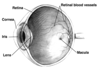
Photo from wikipedia
In this longitudinal study on fovea-off rhegmatogenous retinal detachment, we observed continuous visual acuity improvement and changes of the ellipsoid zone reflection on en face optical coherence tomography for 2… Click to show full abstract
In this longitudinal study on fovea-off rhegmatogenous retinal detachment, we observed continuous visual acuity improvement and changes of the ellipsoid zone reflection on en face optical coherence tomography for 2 years after retinal reattachment surgery. Metamorphopsia did not improve after 12 months, and aniseikonia remained unchanged. Purpose: To investigate the long-term changes in visual function and outer retinal abnormalities on en face optical coherence tomography after fovea-off rhegmatogenous retinal detachment and to assess associations between functional outcomes and outer retinal abnormalities. Methods: Prospective, observational study. The following data were collected at 1, 3, 6, 12, and 24 months after retinal reattachment: Best-corrected visual acuity, metamorphopsia (M-CHARTS), aniseikonia (New Aniseikonia Test), altered ellipsoid zone reflectivity, outer retinal folds, macular detachment demarcation, and subfoveal fluid. Results: Thirty-eight patients were included. Best-corrected visual acuity improved significantly from 1 to 12 months and from 12 to 24 months (P < 0.001; P = 0.022). Vertical and horizontal metamorphopsia improved significantly from 1 to 12 months (P < 0.001; P = 0.002), and at 24 months, scores of ≥0.2° were present in 54% and 42% of patients, respectively. The degree of aniseikonia did not change. Best-corrected visual acuity and aniseikonia scores were positively associated with outer retinal fold (r 0.4, P = 0.009; r 0.4, P = 0.048). A gradual normalization of outer retinal reflectivity took place during 24 months. Conclusion: Visual acuity improved significantly during the second year after reattachment surgery for fovea-off rhegmatogenous retinal detachment, in parallel with normalization of outer retinal abnormalities on en face optical coherence tomography. Metamorphopsia did not improve after 12 months, and aniseikonia remained unchanged.
Journal Title: Retina
Year Published: 2023
Link to full text (if available)
Share on Social Media: Sign Up to like & get
recommendations!