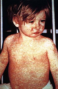
Photo from wikipedia
Purpose: To assess the capability of Scheimpflug-based densitometry of the cornea to quantify light chain deposits in patients with active monoclonal gammopathies. Methods: This is a case–control study in which… Click to show full abstract
Purpose: To assess the capability of Scheimpflug-based densitometry of the cornea to quantify light chain deposits in patients with active monoclonal gammopathies. Methods: This is a case–control study in which data from a leading tertiary university center in myeloma care were analyzed. Ten eyes of 5 patients with monoclonal gammopathy and 26 eyes of 13 healthy controls undergoing clinical evaluation and Scheimpflug-based measurements were included in the study. The main outcome measures were densitometry data of the 4 corneal layers—anterior layer (AL), central layer (CL), posterior layer, and total layer (TL)—in 4 different annuli (central annular zone 0–2 mm, intermediate annular zone 2–6 mm, peripheral annular zone 6–10 mm, and total annular zone 0–12 mm). Results: In 8 eyes of 4 patients with IgG-based gammopathy, corneal light backscatter was highest in the AL and decreased with increasing corneal depth. The peripheral annular zone showed a higher densitometry value compared with the corneal center. Compared with healthy controls, the AL (P < 0.001), the CL (P < 0.001), and the TL (P < 0.001) had significantly higher corneal light backscatter in patients with gammopathy in the total and the peripheral annular zones. In one patient with predominantly IgA-based disease, corneal light backscatter was not elevated. Conclusions: Scheimpflug-based densitometry of the cornea is able to quantify opacification by immunoglobulin G light chain deposits in monoclonal gammopathies. This noninvasive technique can complement presently used in vivo confocal microscopy and corneal photography to objectivize corneal changes. Densitometry might allow monitoring of corneal immunoglobulin deposits in follow-up examinations.
Journal Title: Cornea
Year Published: 2017
Link to full text (if available)
Share on Social Media: Sign Up to like & get
recommendations!