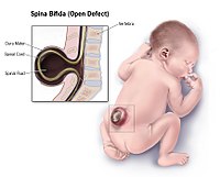
Photo from wikipedia
Introduction: Complete non-mosaic trisomy 22 is a fatal chromosomal disorder that only few fetuses can survive over 12 weeks as reported. Prenatal sonographic findings combined with postnatal or postmortem discoveries… Click to show full abstract
Introduction: Complete non-mosaic trisomy 22 is a fatal chromosomal disorder that only few fetuses can survive over 12 weeks as reported. Prenatal sonographic findings combined with postnatal or postmortem discoveries showed characteristic multi-systematic anomalies. Patient concerns: The unborn baby of a 35-year-old pregnant woman was found to have several anomalies during a prenatal sonographic scan, including intrauterine growth retardation, ventricular septal defect, flat facial profile, and unclear bilateral kidney structures. Diagnoses: The fetus was diagnosed as having complete non-mosaic trisomy 22 by chromosomal analysis. Interventions: The pregnancy was terminated at 24 weeks, and autopsy was permitted. Outcomes: Postmortem examinations revealed additional long-sectional spina bifida occulta and imperforate anus. Conclusions: This was the first time a case of spinal cord defect was reported in trisomy 22 fetuses. More attention should be paid to the spinal cord during sonographic examinations in trisomy 22 fetuses.
Journal Title: Medicine
Year Published: 2018
Link to full text (if available)
Share on Social Media: Sign Up to like & get
recommendations!