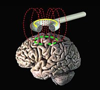
Photo from wikipedia
To investigate the characteristics of pelvic floor surface electromyography parameters on the basis of Glazer assessment in women 42 days postpartum, and to analyze the predictive value of surface electromyography… Click to show full abstract
To investigate the characteristics of pelvic floor surface electromyography parameters on the basis of Glazer assessment in women 42 days postpartum, and to analyze the predictive value of surface electromyography (sEMG) in postpartum stress urinary incontinence. This is a retrospective study. Three thousand twenty-nine females in total who were screened 42 days postpartum in Jinniu District Maternal and Children’s Health Hospital of Chengdu from January 2019 to December 2020 were selected, and were randomly allocated into stress urinary incontinence (SUI) (n = 509) and the non-SUI group (n = 2520). Pelvic floor surface electromyography was performed by the same physiotherapists. The evaluation parameters included the average EMG value in the pre-resting baseline, the maximum sEMG value, the rising time, the descent time in the fast-twitch phase, and the average sEMG value in the slow-twitch phase. Mean value and modifiability of EMG value in post-resting stage. The disparities of the mentioned parameters hereinabove in the SUI and non-SUI groups were made comparison, and the relationship between stress urinary incontinence and sEMG parameters was analyzed by multiple logistic regression analysis. The prevalence of SUI was 16.8% in women 42 days after delivery. Body mass index and vaginal delivery were risk factors for SUI. Among the sEMG parameters of the SUI group and the non-SUI group, the maximum EMG values in the fast-twitch phase (28.81 ± 14.41 vs 30.41 ± 15.15), the rising time in the fast-twitch phase (0.55 ± 0.36 vs 0.51 ± 0.30), and the Phase descent time (0.76 ± 0.76 vs 0.68 ± 0.65), mean slow-twitch phase EMG (17.82 ± 10.10 vs 19.69 ± 15.62), slow-twitch phase variability (0.28 ± 0.12 vs 0.26 ± 0.10), are statistically different (P < .05). In the SUI group, body mass index (estimated parameter = 0.029, P = .023), mean EMG during slow-twitch phase (estimated parameter = -0.013, P = .004) were relevant to stress urinary incontinence after delivery. The sEMG based on Glazer protocol indicates the activity of slow-twitch muscle fibers in SUI patients are decreased, and there is a correlation with the occurrence of stress urinary incontinence. sEMG can be applied as a quantitative evaluation tool of the pelvic floor analysis in postpartum SUI.
Journal Title: Medicine
Year Published: 2023
Link to full text (if available)
Share on Social Media: Sign Up to like & get
recommendations!