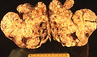
Photo from wikipedia
To the Editor: There is a traditional approach to the surgical excision of mucinous borderline ovarian tumors that includes removal of the appendix in addition to unilateral/bilateral salpingo-oophorectomy and omental… Click to show full abstract
To the Editor: There is a traditional approach to the surgical excision of mucinous borderline ovarian tumors that includes removal of the appendix in addition to unilateral/bilateral salpingo-oophorectomy and omental biopsy with or without hysterectomy. Despite growing evidence that this is an unnecessary procedure (1–5), the practice continues to be widespread. We conducted a single-center, retrospective, observational study of all women diagnosed at The Royal London Hospital with mucinous borderline ovarian tumors between August 2005 andMay 2013. The aim of the study was 3-fold: (i) to establish the number of cases of mucinous borderline ovarian tumors in which the appendix was removed over an 8-yr period, (ii) to determine howmany of those appendices were abnormal, and (iii) to assess whether patients who retained their appendices suffered later sequelae as a result (Fig. 1). Mucinous borderline ovarian tumors account for 30% to 50% of epithelial ovarian borderline tumors and have been previously subdivided into intestinal and endocervical types based on the appearance of their epithelial lining—the former being the most common, and the latter now considered a distinct category renamed ‘‘seromucinous’’ in the current WHO classification scheme (6). They occur across a wide age range encompassing the premenarche, the reproductive period, and the postmenopause. These tumors, the size of which can vary dramatically, are usually unilateral. The presence of bilateral ovarian lesions should prompt greater consideration of metastatic rather than primary disease. The mucinous tumors exist on a spectrum from benign (mucinous cystadenoma) through borderline to malignant (mucinous carcinoma) and each component of this spectrum can exist within the same lesion. Within the borderline category there are 2 further subtypes: mucinous borderline ovarian tumors with marked cytologic atypia are classified as borderline ovarian tumors with intraepithelial carcinoma and those with early stromal invasion (no >5mm) are referred to as borderline tumors with microinvasion. Mucinous borderline tumors are slow-growing and carry an excellent prognosis. They do not present with peritoneal implants and therefore only exist as stage I disease. Limited reports of recurrence and metastatic disease do exist but it is likely that inadequacies of treatment (cystectomy rather than salpingo-oophorectomy) and tissue sampling account for these aberrant descriptions (7,8). Eighty-one women were diagnosed with a mucinous borderline ovarian tumor during the study period, of which 41 underwent appendicectomy. Of the 41 appendicectomies, 37 were performed as part of the primary surgery and 4 as a separate completion staging procedure following a postsurgical histologic diagnosis of mucinous borderline ovarian tumor. Forty women, therefore, retained their appendices. Of the 41 appendices that were removed, none showed any gross abnormality or histologic evidence of neoplasia. All were reported as being within normal limits. Among the group of women who did not undergo appendicectomy, none suffered later complications related to appendiceal pathology. Historically, the appendix was removed during surgery for mucinous ovarian neoplasms because of perceived difficulties in identifying the primary site of the tumor. It was believed that what was ostensibly a mucinous ovarian neoplasm may, in fact, represent metastasis from an occult appendiceal lesion. However, greater understanding of mucinous neoplasia and advances in immunohistochemistry have improved diagnostic clarity (9,10). Pseudomyxoma peritonei (PMP) requires separate consideration. This is a form of tumor implantation whereby glandular epithelial cells are deposited in the peritoneal cavity, producing large volume mucinous ascites. Originally, it was thought that these cells were derived from mucinous borderline or malignant neoplasms of the appendix, ovary, or pancreas. 81 Women
Journal Title: International Journal of Gynecological Pathology
Year Published: 2018
Link to full text (if available)
Share on Social Media: Sign Up to like & get
recommendations!