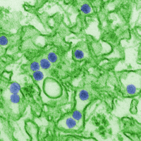
Photo from wikipedia
Abstract In congenital Zika virus syndrome (CZS), the most frequent radiological findings are calcifications in the cortical–white matter junction and malformations of cortical development (pachygyria or polymicrogyria, which occur predominantly… Click to show full abstract
Abstract In congenital Zika virus syndrome (CZS), the most frequent radiological findings are calcifications in the cortical–white matter junction and malformations of cortical development (pachygyria or polymicrogyria, which occur predominantly in the frontal lobes, or a simplified gyral pattern), ventriculomegaly, enlargement of the cisterna magna and the extra-axial subarachnoid space, corpus callosum abnormalities, and reduced brain volume. This syndrome can also result in a decrease in the brainstem and cerebellum volumes and delayed myelination. Infants with CZS may show venous thrombosis and lenticulostriate vasculopathies. Over a 3-year follow-up period, many infants with CZS showed hydrocephalus, reduction in brain calcifications, and greater reduction in brain thickness.
Journal Title: Topics in Magnetic Resonance Imaging
Year Published: 2019
Link to full text (if available)
Share on Social Media: Sign Up to like & get
recommendations!