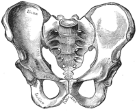
Photo from wikipedia
Introduction The medial thighplasty is a procedure where patients may attain superior mobility, hygiene, and cosmesis. Most surgeons use attachment of the superficial fascial system (SFS) of the thigh flap… Click to show full abstract
Introduction The medial thighplasty is a procedure where patients may attain superior mobility, hygiene, and cosmesis. Most surgeons use attachment of the superficial fascial system (SFS) of the thigh flap to the Colles fascia, whereas others attach the SFS to the pubic ramus periosteum. Because of a high complication profile, we aim to elucidate the clinical, biomechanical, and anatomic qualities of the Colles fascia versus the pubic ramus periosteum. Materials and Methods We performed a 17-year retrospective review documenting clinical complications, a biomechanical analysis of sutures placed in different tissue layers of the thigh, and a histologic analysis surrounding the ischiopubic ramus. Separate suture pull-out strength testing was conducted on cadaveric tissue using an Admet MTEST Quattro with no. 1 Vicryl suture and tissue grips at a displacement rate of 2.12 mm/s. Simultaneous displacement and force were acquired at 100 Hz and with measurements obtained at regular intervals between the pubic symphysis and the ischial tuberosity in both the Colles fascia and the deeper periosteal layers of the thigh. A histologic analysis was performed at 3 points along the ischiopubic ramus using paraffin-embedded large mount tissue sections stained with hematoxylin, eosin, and Gomori trichrome. Results Thirty-nine patients underwent medial thighplasty with a 46.16% complication rate. Suture pull-out force of the suspected superficial Colles fascia sites was, on average, 72.8% less than values from the deeper periosteum tissue. Anchor points in the Colles fascia elongated 17.4% further before failure than those in the periosteum. There was noticeable variability between anchor points and across samples. The histologic sections suggest that the Colles fascia from the different regions of the ischiopubic ramus varies considerably in both continuity and collagen fiber content with no discernible pattern. The periosteal and muscular fascial layers were more continuous histologically with direct attachments into the pubis and ischium. Conclusions Anchoring of the SFS to the periosteum did not improve our complication profile when compared with the literature. Both the biomechanical and histologic analyses demonstrate that the Colles fascia is highly variable in organizations with coincident variability in tissue strength. Our results require further study to identify the optimal surgical technique for medial thighplasty.
Journal Title: Annals of Plastic Surgery
Year Published: 2017
Link to full text (if available)
Share on Social Media: Sign Up to like & get
recommendations!