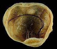
Photo from wikipedia
CLINICAL REPORT A 5-year-old female child was presented to us with a history of painless swelling in the lower part of neck; noticed by mother since 15 days. On examination,… Click to show full abstract
CLINICAL REPORT A 5-year-old female child was presented to us with a history of painless swelling in the lower part of neck; noticed by mother since 15 days. On examination, we found a nodular swelling of size 1 cm 0.8 cm in the supra sternal notch (Fig. 1A); the lower limit of the swelling was not clearly appreciated. The swelling did not move with cough, swallowing, or on protrusion of tongue. Child was evaluated with sonography, which revealed a hypoechoic swelling confined to supra sternal region (Fig. 2E). To rule out any other anterior mediastinal pathology, we did contrast-enhanced computed tomography of chest (CECT), which revealed, a solitary hypo dense mildly enhancing benign cystic lesion without any calcification in the supra sternal region without any other pathology in the neck as well as in the thorax (Fig. 2F-H). For definitive diagnosis, child underwent excisional biopsy under general anesthesia. During the procedure, we found soft cystic nodular swelling of 1.2 cm 0.8 cm, with white pultaceous material in it. Histopathology confirmed it to be epidermoid cyst lined by keratinized stratified squamous epithelium with keratin debris in the lumen of the cyst without any adnexal structures (Fig. 1D). Child is doing well at one year’s follow-up.
Journal Title: Journal of Craniofacial Surgery
Year Published: 2020
Link to full text (if available)
Share on Social Media: Sign Up to like & get
recommendations!