Photo from wikipedia
A 62-year-old woman presented to our neuroophthalmology clinic in referral from her comprehensive ophthalmologist. Past medical and ocular history included diverticulosis, depression, and early nonexudative age-related macular degeneration, which had… Click to show full abstract
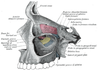
A 62-year-old woman presented to our neuroophthalmology clinic in referral from her comprehensive ophthalmologist. Past medical and ocular history included diverticulosis, depression, and early nonexudative age-related macular degeneration, which had been diagnosed approximately 1 year before presentation. She endorsed “dimmer” vision on the left compared with the right for the past year. For the past 6 months, she had noticed slowly progressive “blind spots” in her vision in the left eye, which she noted with routine Amsler grid monitoring. Initial exam in our clinic demonstrated mildly decreased visual acuity on the left, full extraocular movements bilaterally, and mild left optic disc pallor. A 0.6 log unit relative afferent pupillary defect (RAPD) was noted. Ancillary testing included Humphrey 30-2 visual field testing which revealed full visual fields on the right and relatively nonspecific changes in the left eye, and optical coherence tomography (OCT) of both macula and optic nerves. The retinal ganglion cell layer thickness was normal bilaterally, with mild temporal circumpapillary thinning in 2 clock-hours on the left eye. MRI revealed a T2-hyperintense, heterogeneously enhancing, 1.5· 1-cm extraconal mass of the left orbital apex which extended through the inferior orbital fissure and into the pterygopalatine fossa (Fig. 1). The left optic nerve was displaced superiorly with compression against the roof of the optic canal. Computed tomography of the orbit was obtained which demonstrated homogeneous hyperintensity of the mass with associated bony remodeling of the inferior orbital fissure and medial orbital wall (Fig. 2). An endoscopic approach through the maxillary and sphenoid sinuses was deemed preferable to an orbital
Journal Title: Journal of Neuro-Ophthalmology
Year Published: 2021
Link to full text (if available)
Share on Social Media: Sign Up to like & get
recommendations!