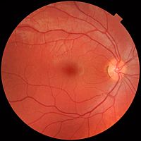
Photo from wikipedia
Glaucoma is a chronic progressive optic neuropathy that causes visual impairment or blindness if left untreated. It is crucial to diagnose it at an early stage in order to enable… Click to show full abstract
Glaucoma is a chronic progressive optic neuropathy that causes visual impairment or blindness if left untreated. It is crucial to diagnose it at an early stage in order to enable treatment. Fundus photography is a viable option for population-based screening. A fundus photograph enables the observation of the excavation of the optic disk—the hallmark of glaucoma. The excavation is quantified as a vertical cup-to-disk ratio (VCDR). The manual assessment of retinal fundus images is, however, time-consuming and costly. Thus, an automated system is necessary to assist human observers. We propose a computer-aided diagnosis system, which consists of the localization of the optic disk, the determination of the height of the optic disk and the cup, and the computation of the VCDR. We evaluated the performance of our approach on eight publicly available datasets, which have, in total, 1712 retinal fundus images. We compared the obtained VCDR values with those provided by an experienced ophthalmologist and achieved a weighted VCDR mean difference of 0.11. The system provides a reliable estimation of the height of the optic disk and the cup in terms of the relative height error (RHE = 0.08 and 0.09, respectively). The Bland–Altman analysis showed that the system achieves a good agreement with the manual annotations, especially for large VCDRs which indicate pathology.
Journal Title: IEEE Access
Year Published: 2019
Link to full text (if available)
Share on Social Media: Sign Up to like & get
recommendations!