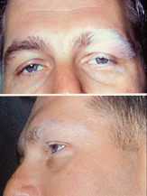
Photo from wikipedia
Inflammatory eye diseases, such as uveitis, scleritis and retinal vasculitis, are among the most common extra‐articular manifestations of rheumatic disease and systemic vasculitis. Uveitis is the most frequent ocular complication… Click to show full abstract
Inflammatory eye diseases, such as uveitis, scleritis and retinal vasculitis, are among the most common extra‐articular manifestations of rheumatic disease and systemic vasculitis. Uveitis is the most frequent ocular complication of rheumatic diseases and is a major cause of severe visual impairment. It affects people of all ages and varies significantly by geographic location and age of the patient. The incidence of uveitis is 25 cases per 100 000 persons with a prevalence rate of 58 per 100 000 persons.1 Uveitis is a generic term used to describe inflammation of the uvea, which is composed of the iris, ciliary body, and choroid. However, any area of the eye can be involved as the inflammatory response often spills over to involve contiguous or adjacent ocular tissues such as the cornea, anterior chamber, vitreous and retina. Uveitis can be subdivided into anterior, intermediate, posterior, and panuveitis based on the primary anatomical location of the inflammation in the eye. Anterior uveitis occurs when the anterior segment (iris and ciliary body) is the predominate site of inflammation. Intermediate uveitis is defined by inflammation of the vitreous cavity and pars plana, while posterior uveitis involves the retina and choroid. Panuveitis involves inflammation of the entire uveal tract.2 Uveitis is most commonly idiopathic or undifferentiated, although it can be associated with rheumatic, autoimmune, inflammatory or infectious systemic diseases, or be traumatic, or drug‐induced (eg rifabutin, check point inhibitors and bisphosphonates). Patients with uveitis may present with concurrent systemic symptoms or infectious diseases that suggest an etiology affecting more than just the eye, or with isolated eye symptoms. Idiopathic uveitis accounts for 48% to 70% of cases.3 Uveitis is generally non‐ infectious (up to 85%) and anterior (80%).2 Systemic inflammatory disorders commonly associated with anterior uveitis include human leukocyte antigen (HLA)‐B27‐associated entities, juvenile idiopathic arthritis, inflammatory bowel disease, sarcoidosis, Behçet's disease or tubulo‐interstitial nephritis (TINU). Multiple sclerosis, sarcoidosis, and TINU may be associated with intermediate uveitis, while Vogt‐ Koyanagi‐Harada (VKH) syndrome, systemic lupus erythematosus, Behçet's disease, and sarcoidosis can cause posterior or panuveitis. Masquerade syndromes associated with lymphoma and leukemia can present with posterior or panuveitis. Infectious diseases account for approximately 15% of all cases of uveitis. These include viruses (herpes simplex, varicella zoster, cytomegalovirus), bacteria (streptococcal/ staphylococcal endophthalmitis, syphilis, tuberculosis, bartonella, Lyme borreliosis etc), or parasites/worms (toxoplasmosis, toxocara, nematodes) or other atypical infections.4 Different patterns of uveitis are encountered around the world due to the influence of many factors including genetic, ethnic, geographic and environmental factors. Such factors have a major impact on the causes and clinical phenotypes of uveitis and have been identified in a number of epidemiology studies. As examples, VKH and sarcoidosis are common in Japan, Behçet's disease in the Mediterranean and Asia, tuberculosis‐associated uveitis in India5 and toxoplasmosis in South America.6-8 Over the past 15 years, the Standardization of Uveitis Nomenclature (SUN) Working Group, an international group of 79 uveitis experts from 18 countries and 62 clinical centers, has developed an anatomic classification for uveitis. This includes descriptors for uveitis, severity gradings for anterior chamber cells and flare, and definitions for uveitis activity.9 The SUN group is in the final stages of developing classification criteria for 25 clinical phenotypes of uveitis which will hopefully standardize reporting and classification of these entities.10
Journal Title: International Journal of Rheumatic Diseases
Year Published: 2019
Link to full text (if available)
Share on Social Media: Sign Up to like & get
recommendations!