Photo from wikipedia
Endoscopic ultrasound-guided fine needle aspiration (EUS-FNA) is an accurate and minimally invasive modality to obtain samples for cytological and biochemical analysis. As this modality is being used with increasing frequency,… Click to show full abstract
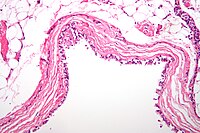
Endoscopic ultrasound-guided fine needle aspiration (EUS-FNA) is an accurate and minimally invasive modality to obtain samples for cytological and biochemical analysis. As this modality is being used with increasing frequency, it is essential to analyse its safety profile and timing of complications. Here, we report a case of intraabdominal abscess presenting 45 days after EUS-FNA, requiring antibiotics and surgical debridement. A 65-year-old woman presented to clinic with a 5-year history of epigastric pain, on a background of diabetes, obesity, obstructive sleep apnoea and hypertension. Transabdominal ultrasound demonstrated a 6-cm mass in the head of the pancreas. This was not seen on an ultrasound performed 3 years previously. Computerized tomography (CT) scan demonstrated a 6.3 × 5.9 cm cystic mass abutting the inferior aspect of the pancreatic head and transverse portion of the duodenum. This was thought to represent a mucinous cystic neoplasm of the pancreas. Tumour markers (CEA, CA19-9) were normal. As the diagnosis was unclear, EUS-FNA was performed. This demonstrated a solid tumour with cystic degeneration, which appear separate to the stomach and pancreas. FNA was performed with a 22-gauge needle, demonstrating acellular debris consistent with cyst content but no cells that indicated a cystic neoplasm. The overall impression was of a stromal tumour or duplication cyst. While waiting for clinical follow-up, the patient presented to the emergency department 45 days after EUS-FNA with generalized abdominal pain associated with fevers, nausea and vomiting. CT scan of the abdomen (Fig. 1) showed the lesion in the upper abdomen had increased in size and contained multiple pockets of gas with adjacent inflammatory change. She was admitted for a course of intravenous antibiotics. One week after discharge, she represented with similar pain, and repeat CT scan showed increased inflammatory change on the right side of the abdomen. She proceeded to an exploratory laparotomy, where a large abscess was found, associated with a cystic lesion arising from the third portion of the duodenum (Fig. 2). The lesion shared a wall with the duodenum, but there was no obvious communication with the duodenal lumen. The contents of lesion were a thick, creamy paste, similar to that seen in an epidermal cyst. The lesion appeared consistent with a duplication cyst. Cultures from the abdominal fluid grew Streptococcus mitis. Histology confirmed a duodenal duplication cyst lined by keratinizing squamous epithelium. The patient made an uneventful postoperative recovery, and was discharged after 15 days. To our knowledge, this is the first reported case of infection in a duodenal duplication cyst after EUS-FNA, and the longest interval between FNA and presentation with infection (45 days). The risk of infection after EUS-FNA is low and comparable with that of diagnostic endoscopy. In a systematic review, the pooled infection rate after EUS-FNA was 0.046%, with infection of cystic lesions being higher at 0.22%. Accordingly, prophylactic antibiotics are not recommended for FNA of solid lesions, while antibiotic use for FNA of cystic lesions remains controversial because of the slightly higher risk of infection. Duplications of the gastrointestinal tract are rare congenital malformations. It is most commonly seen in distal ileum, followed by the oesophagus and duodenum. It comprise of 2% to 12% of all duplications. Most are diagnosed in early childhood, but 38% of patients are diagnosed after the age of 20 years. As shown in the above case, duodenal duplication cysts are usually located at the second or third part of the native duodenum and closely associated with the pancreatobiliary duct system.
Journal Title: ANZ Journal of Surgery
Year Published: 2017
Link to full text (if available)
Share on Social Media: Sign Up to like & get
recommendations!