Photo from wikipedia
A 12yearold female patient visited our ophthalmology department in 2014 due to an abnormality seen by the optometrist. We found that the visual acuity in both of her eyes was… Click to show full abstract
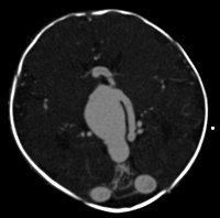
A 12yearold female patient visited our ophthalmology department in 2014 due to an abnormality seen by the optometrist. We found that the visual acuity in both of her eyes was 6/5; her visual fields were intact; and optical coherence tomography (OCT) of both eyes was normal (Figure 1). However, during fundal imaging of the right eye, a novel and incidental occurrence of retinal racemose hemangioma (RRH) was found. RRH is a rare congenital and nonhereditary anatomical arteriovenous malformation with less than 100 cases reported in the literature. They are often diagnosed by ophthalmoscopy; however, f luorescein angiography may be utilised to detect smaller lesions. Ultrasound and OCT may also be used to further confirm the diagnosis (So et al., 2021). Based on the contemporary classification, the patient had Group 2 RRH as shown in Figure 2 on fundal photography and Optos imaging of the patient's right eye, with the left eye imaging included for comparison. RRH is typically unilateral and is divided into 3 groups based on the size and location of the vascular malformation as shown schematically in Figure 3. Group 1 RRHs have an abnormal capillary plexus between the major artery and vein of the arteriovenous malformation and can be difficult to detect. In Group 2, the arteriovenous malformation lacks an intervening capillary bed between the artery and vein. And in Group 3, RRHs present with extensive arteriovenous malformations with dilated and tortuous vessels with difficulty in distinguishing between arteries and veins. Overall, the variation in RRH is wide and can range from a single welldemarcated anastomosis in one retinal quadrant to tumourlike masses with dilated arteries and veins (Vishal et al., 2018). Further, this disease falls under the umbrella of Group 2 of the cerebrofacial arteriovenous metameric syndrome (CAMS) spectrum of arteriovenous malformations (AVMs). Group 2 CAMS AVMs involve the cortex, diencephalon, optic chasm, optic nerve, retina, sphenoid, maxilla and cheek (So et al., 2021). An important differential diagnosis to consider is retinal capillary hemangioma (RCH) found in Von Hippel– Lindau (VHL) disease where up to half of cases are bilateral, and most cases are associated with a prominent feeder vessel. Importantly, RCH lesions appear as welldemarcated round orange to red retinal lesions in the juxtapapillary, midperipheral, superotemporal periphery or inferotemporal periphery of the retina (Turell & Singh, 2010). An example of an RCH is shown in Figure 4, and when compared against scientific literature and the RRHs in Figures 1 and 2, it can be appreciated that RRHs tend not to have a prominent feeder vessel. However, it should be mentioned that dilated vessels are seen in many RRHs (Vishal et al., 2018), and this may pose challenges to distinguish it from RCH. Furthermore, when thinking of differentiators for RRH and RCH, though renal cell carcinoma is the leading cause of death in VHL, kidney pathology is seen in both WyburnMason syndrome and VHL; therefore, investigating kidney disease does not serve as a great differentiator. Genetic testing for the VHL gene mutation is considered a superior diagnostic tool for clinicians to increase confidence that any anatomical variation found is that of RRH rather than of RCH, which is inherited in an autosomal dominant fashion and has detection rates as high as 99% on tests (Turell & Singh, 2010). Optical coherence tomography can be used to follow changes in the nerve fibre layer, macula and retina over time. It is also useful for diagnosing macular oedema and serous retinal detachment (So et al., 2021). Since diagnosis, our patient remained asymptomatic until 2019, after which the OCT started to detect the mild onset of leakage surrounding the RRH superior to the macula in the form of intraretinal fluids (Figure 5). However, we were not particularly alarmed as the leaking of fluid did not Received: 8 July 2022 | Accepted: 14 August 2022
Journal Title: Acta Ophthalmologica
Year Published: 2022
Link to full text (if available)
Share on Social Media: Sign Up to like & get
recommendations!