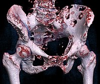
Photo from wikipedia
We read the recently reported study by Ujjani et al (2016) with interest. Their retrospective study evaluated the diagnostic value of F-fluoro-2-deoxy-D-glucose positron emission tomography (FDG-PET) in identifying bone marrow… Click to show full abstract
We read the recently reported study by Ujjani et al (2016) with interest. Their retrospective study evaluated the diagnostic value of F-fluoro-2-deoxy-D-glucose positron emission tomography (FDG-PET) in identifying bone marrow involvement in various lymphoma subtypes using bone marrow biopsy (BMB) as reference standard. Focally or diffusely increased bone marrow uptake at FDG-PET was observed in 6/10 (60%) patients with diffuse large B-cell lymphoma (DLBCL) and positive BMB, 24/35 (68 6%) patients with follicular lymphoma and positive BMB and 3/3 (100%) patients with Hodgkin lymphoma and positive BMB (Ujjani et al, 2016). Most of the patients in whom FDG-PET failed to identify bone marrow involvement already had advancedstage disease according to imaging, and bone marrow status would not have changed treatment strategy in these patients. Ujjani et al (2016) concluded FDG-PET to be highly accurate for detecting bone marrow involvement in DLBCL and Hodgkin lymphoma, and that BMB may not be necessary if the patient is already considered to have advanced-stage disease by FDG-PET imaging. Although these conclusions sound very promising, several comments have to be made. First, we do not agree with their claim that FDG-PET is highly accurate in identifying bone marrow involvement in DLBCL patients. A metaanalysis reported FDG-PET to have a high patient-based sensitivity for the detection of bone marrow involvement in DLBCL, ranging between 70–95% (Adams et al, 2014). However, all studies included in that meta-analysis used both BMB and follow-up FDG-PET scans as reference standard (Adams et al, 2014). Although a positive BMB is considered proof of bone marrow involvement, decrease in bone marrow FDG uptake following treatment may not be sufficient proof of baseline bone marrow involvement. Thus, the sensitivity of FDG-PET is probably lower than documented in that meta-analysis (Adams et al, 2014). This statement is supported by the results reported by Ujjani et al (2016), in which almost half (4/10) of their DLBCL patients with bone marrow involvement according to BMB did not have focally or diffusely increased bone marrow FDG uptake at PET. Note that the suboptimal sensitivity of FDG-PET to detect (concordant and discordant) bone marrow involvement has also been demonstrated by several other (recent) studies (Adams et al, 2015a) that were not discussed by Ujjani et al (2016). The aforementioned findings clearly underline that the claim of current guidelines (Barrington et al, 2014), that FDG-PET is highly sensitive for the detection of bone marrow involvement in DLBCL, is not supported by scientific evidence and requires rectification. Second, Ujjani et al (2016) did not discuss that, whilst bone marrow involvement according to BMB has been established as a major adverse prognostic factor in DLBCL (Sehn et al, 2011; Adams et al, 2015a), this is not the case for bone marrow involvement as determined by FDG-PET. Indeed, the majority of studies on this topic showed bone marrow status at FDG-PET to have no prognostic value at all (Adams et al, 2015a). Consequently, the recently published National Comprehensive Cancer Network Internal Prognostic Index (NCCN-IPI) does not incorporate imaging-based bone marrow involvement, but only biopsy proven bone marrow involvement for risk stratification (Zhou et al, 2014). Third, it was not reported how many patients with diffusely increased bone marrow FDG uptake were observed, and how these findings correlated to the bone marrow histology. Note that the interpretation of diffusely increased bone marrow uptake is not well established and appears to vary considerably among different lymphoma subtypes (Adams et al, 2015a,b). Fourth, although most of the patients with Hodgkin lymphoma, DLBCL and follicular lymphoma in which FDG-PET failed to identify bone marrow involvement already had advanced-stage disease at FDG-PET, it was not determined how many of these patients were already classified as advanced-stage disease, as determined by computed tomography (CT) only. Consequently, the additional value of FDG-PET to CT in terms of treatment planning was not assessed. Note that BMB has been reported to have no consequences in terms of treatment planning in large proportions of patients even if an FDG-PET based evaluation of the bone marrow is not performed (Adams et al, 2015c). Fifth, although bone marrow involvement missed by FDGPET at baseline did not result in a major alteration in patient management in terms of treatment planning, the suboptimal sensitivity of FDG-PET to detect bone marrow involvement in non-Hodgkin lymphomas clearly underlines that a negative FDG-PET scan at end-of-treatment cannot exclude residual lymphomatous deposits in the marrow. This may (partially) explain why a high proportion of DLBCL patients in ‘complete remission’ according to endof-treatment FDG-PET develops disease relapse during correspondence
Journal Title: British Journal of Haematology
Year Published: 2017
Link to full text (if available)
Share on Social Media: Sign Up to like & get
recommendations!