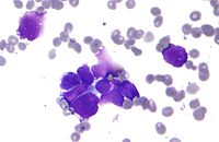
Photo from wikipedia
Three months later, the man underwent follow-up flexible cystoscopy in the outpatient clinic. At this point, a voluminous necrotic excrescency was noted, growing from the right side of the bladder… Click to show full abstract
Three months later, the man underwent follow-up flexible cystoscopy in the outpatient clinic. At this point, a voluminous necrotic excrescency was noted, growing from the right side of the bladder where the previous primary tumour had been resected. In addition to this finding, it was noted that a papillomatous protuberance was emerging under a layer of fibrin in the bladder dome as well as from the left side of the bladder wall around a diverticulum. Resection of the necrotic excrescency and biopsies were undertaken via TURBT 1 week later. Radical excision was not feasible and several small pathological areas remained after the TURBT. Histopathological examination of the resected tissue from the right bladder wall showed papillomatosis and welldifferentiated, acanthotic squamous epithelium with marked hyperkeratosis. No irregular invasive nests were found, but a ‘pushing border’ was observed in the specimens. The tumour was categorized as suspicious for verrucous carcinoma (VC; Fig. 1). The second biopsy sampled around the diverticulum showed papillary hyperplasia with keratinizing squamous metaplasia. Immunoreactivity for Ki67 indicated high proliferative activity in both specimens. The specimens were revised at a specialized tertiary university hospital because of the unusual findings. The suspicion of VC, however, remained. The patient had no history of travelling, and no Schistosoma haematobium eggs were identified in the specimens.
Journal Title: BJU International
Year Published: 2022
Link to full text (if available)
Share on Social Media: Sign Up to like & get
recommendations!